Structural MRI
Structural MRI is used to visualize and measure the 3D anatomy of the human brain. The MRI signal used to produce structural images is sensitive to the amount of water in different types of tissue. Tissues with high signal appear bright white, while areas with low signal appear dark. There are different kinds of structural MRI images. At the CCBBI we commonly collect T1-weighted and T2-weighted images.
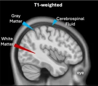
T1-weighted images highlight areas with low water concentrations, such as white matter (red marker). Areas with high water concentration, such as gray matter and cerebrospinal fluid (blue markers) appear dark.
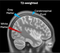
T2-weighted images highlight areas with high water concentrations, such as gray matter and cerebrospinal fluid (blue markers). Areas with low water concentrations, such as white matter (red markers) appear dark.