Research Day 2021
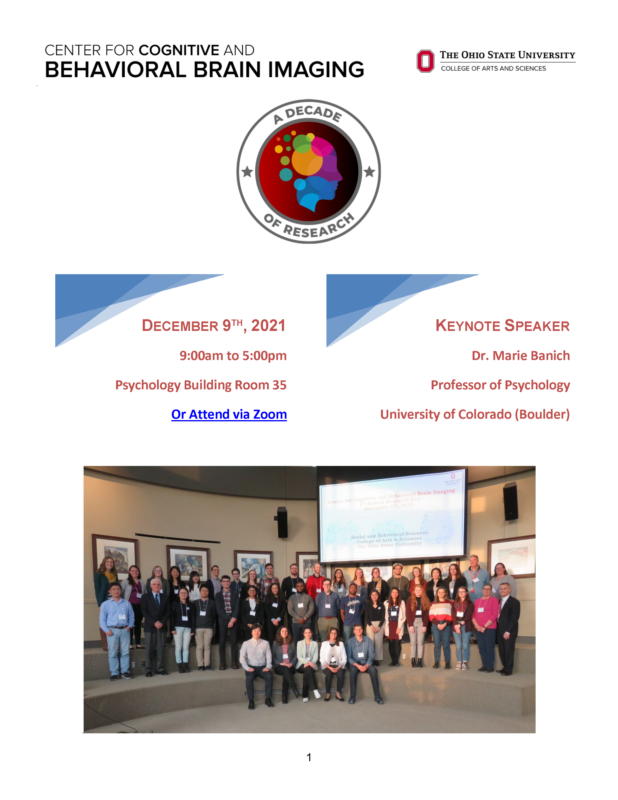
CCBBI Research Day Agenda - December 9, 2021
9:00 am Registration and poster setup
9:45 am Opening Remarks: Dr. Ruchika Prakash (CCBBI Director)
10:00 am Keynote Talk: Dr. Marie Banich, University of Colorado (Boulder)
11:00 am Featured Faculty Presentations
12:30 pm Lunch
1:45 pm Graduate Student Oral Presentations
3:15 pm Poster Flash Talks
3:45 pm Poster Presentations and Refreshments
4:45 pm Closing Remarks
Keynote Address: Dr. Marie Banich

Dr. Marie Banich is a professor of Psychology at the University of Colorado (Boulder), and she also serves as the Executive Director of both the Institute of Cognitive Science and the Intermountain Neuroimaging Consortium. You may also recognize Dr. Banich for her work as the author of the Cognitive Neuroscience textbook—a text, now in its 4th edition, that many of us have encountered along the way in our academic journeys.
Title: "Stop thinking about it”! Neural and cognitive mechanisms for actively removing information from working memory
Abstract: How can we as scientists determine when someone has stopped thinking of something? Said differently, how can we find an experimental signature of a thought that no longer exists? In this talk I will discuss behavioral and neuroimaging research that addresses this question to elucidate the cognitive control mechanisms that allow information in working memory to be actively removed from the current focus of attention. Most paradigms used in cognitive psychology and cognitive neuroscience to study working memory do not actually require information to be removed nor do they measure when such removal has occurred. In contrast, our work using a marriage of functional neuroimaging and machine learning techniques, including multi-voxel pattern analysis (MVPA), in conjunction with behavioral experiments has been able to verify that a thought has indeed been removed. Our work provides evidence that there are at least three distinct ways of removing information from working memory: by replacing it with something else, by specifically targeting it for suppression, and by clearing the mind of all thought. In this talk, I will discuss a) the neural mechanisms that enable each of these three types of operations, b) provide evidence regarding the time course of the removal of information from working memory, and c) elucidate the consequences of these removal operations for the encoding of new information, which is critical for new learning. In addition to having implications for enhancing our knowledge of the cognitive control operations that work on information in working memory, I will also discuss the implications of this work for psychological and psychiatric disorders, many of which are characterized by recurrent or intrusive thoughts that individuals cannot remove from the current focus of attention.
Featured Faculty Presenters

Lei Wang, PhD
Professor, Department of Psychiatry and Behavioral Health
Deep Brain Structural Shape in Schizophrenia

Dylan Wagner, PhD
Associate Professor, Department of Psychology
There are people living inside your brain: How similarities in neural representation underlie similarities in person perception

Stephanie Gorka, PhD
Assistant Professor, Department of Psychiatry and Behavioral Health
Brain-Behavior Sensitivity to Uncertainty as a Novel Transdiagnostic Treatment Target

Ruchika Prakash, PhD
Professor, Department of Psychology, CCBBI Director
Towards Brain-Based Mechanistic and Endpoint Markers of Treatment Gains
Graduate Student Oral Presentations
Visual distraction disrupts category-tuned attentional filters in ventral visual cortex
Lasyapriya Pidaparthi, Blaire Dube, & Julie D Golomb
Keywords: attentional capture, attentional filtering, fMRI, category-selective regions, distraction, visual search
To compensate for the complexity of our visual environments, we rely on attention to selectively filter goal-relevant information from irrelevant information. During visual search, for instance, attention prioritizes target-matching information in the environment. However, if distracting information appears during this search, spatial attention can be captured, prolonging search. Does distraction also disrupt feature- or category-based attentional filters that prioritize task-relevant information? To examine this, we measured the neural signatures of attentional filtering specific to object category using human fMRI. In this experiment, participants were presented with 2x2 arrays of hybrid face-house images; they performed a 1-back task based on the designated stimulus category (attend-faces, attend-houses blocks), with the target item’s location indicated by a solid white border. On some trials, a salient distractor (a dotted white border) appeared briefly around one of the nontarget locations. On distractor-absent trials, we found the predicted pattern of attentional modulation: greater BOLD activation in the fusiform face area (FFA) during attend-faces blocks and in the parahippocampal place area (PPA) during attend-houses blocks, illustrating the efficacy of attentional filtering for the target category. On distractor-present trials, however, we observed a relative increase in activation reflecting the processing of the nontarget category (i.e., increased FFA activation during attend-houses and increased PPA activation during attend-faces), suggesting a disruption of the attentional filter selective for object category. This disruption was robust and global throughout the visual scene, resulting in the errant processing – and early on, prioritization – of the irrelevant category from across the entire visual scene. These results demonstrate a novel consequence of attentional capture and provide direct evidence for the Filter Disruption Theory: in addition to disrupting spatial attention, distraction also disrupts global, nonspatial attentional filters tuned to goal-relevant information.
The boundary between real and fictional others within the medial prefrontal cortex is blurred in lonelier individuals
Tim Broom & Dylan Wagner
Keywords: fictional characters; fMRI; narrative; loneliness; representational similarity analysis
The social surrogacy hypothesis suggests that for individuals whose belongingness needs are unmet, fictional characters can stand in for friends and provide the experience of belonging. In the present study, we investigated how loneliness may modulate neural representations of real friends and fictional ones. Fans of the HBO television series Game of Thrones performed a common trait-evaluation task for the self, 9 real-life friends/acquaintances, and 9 fictional characters from Game of Thrones while undergoing functional magnetic resonance imaging. Using representational similarity analysis to examine neural pattern similarity between pairs of identities within the medial prefrontal cortex (MPFC), we found evidence of distinct boundaries between real and fictional others. However, among lonely individuals, the boundary between these two categories was blurred. Similarly, when examining neural similarity between the self and real-life friends/acquaintances and the self and fictional characters, we found that lonelier individuals again showed a blurring of boundaries such that there was increased self-other neural similarity with fictional characters and decreased self-other neural similarity with real others. Together, these results suggest that lonelier individuals may turn to fictional characters to meet belongingness needs that are not met by members of their real-world social networks and that they in turn encode these fictional characters more similarly to friends as well as to the self.
A personalized cortical atlas, generated from individual subject voxelwise connectivity
M. Fiona Molloy & David E. Osher
A major goal for neuroscientists involves mapping the human brain into meaningful units. An ideal atlas provides consistency across studies and populations, corresponds to meaningful organization of cognitive processing, and captures individual differences. Functional connectivity, or co-fluctuations in the fMRI signal, is a particularly promising candidate for defining atlases because of its ability to capture individual differences and predict behavior, brain disorders, and cognition. While many individualized atlases based on functional connectivity have been proposed, there is no single correct solution. Further, it remains unclear how these atlases relate to an individual’s functional regions of interest. Here, we describe a more flexible approach to determine individual solutions across a wide range of resolutions. To construct and validate these personalized atlases, we utilized a large sample of young adults (N= 1,018) with resting state and task fMRI from the Human Connectome Project (van Essen et al. 2012). Group-level parcellations were defined from the average high-resolution connectome of a subset of 500 individuals using k-means clustering along a sequence of networks from 2 to 200. Next, the individualized parcellations were defined for the remaining 518 individuals using a k-nearest neighbors approach informed by the group solutions. We found these personalized atlases: 1. Conserve group-level aspects, 2. Replicate broadscale organization of previously released atlases, and 3. Are stable within individuals. Next, we identified resting state parcels that corresponded to an individual’s task-based activation for motion, working memory, and high-level vision. These parcels resembled an individual’s task-based activity, particularly with motor localizers. Specificity was also observed in motor, place, body, and most face localizers. Overall, we found this parcellation methodology replicates previous group-level results, captures individual differences in functional connectivity at rest, and resembles an individual’s task-based activity. These results echo previous findings on the importance of individual differences and describes a potential method to individually tune previously released group-level parcellations. Further, these results could provide a way to identify, or constrain, individual localizers from resting state data alone. Future work is necessary to decrease the computational load of these analyses, and to test the generalizability of these parcellations to other data and different cognitive tasks.
Gaze-centered spatial representations in human hippocampus
Zitong Lu, Anna Shafer-Skelton, & Julie D. Golomb
As we move our eyes around the world, we can integrate visual input and achieve stable perception across eye movements. However, previous studies have found that our visual system, from primary visual cortex to higher level visual regions, represents object locations in retinotopic (eye-centered) but not spatiotopic (world-centered) coordinates. Is spatiotopic location represented elsewhere in the brain? The hippocampus is also known to be necessary in visual, spatial, and navigational processing. However, how hippocampus might represent object location across eye movements is still unexplored. In this study, we manipulated fixation and stimulus locations in an object perception task and used functional fMRI to record participants’ brain activity. Here, we applied both correlation-based multi-voxel pattern analysis (MVPA) and representational similarity analysis (RSA) to explore the representation of object location and investigate what role does the human hippocampus play in visual stability. We constructed several coding models corresponding to different types of spatial coding patterns and compared them with brain representation patterns from LOC, PPA and hippocampus. Our results found significant retinotopic information instead of spatiotopic information not only in LOC and PPA (consistent with prior findings), but also in hippocampus. Although the strength of the retinotopic representation decreased from LOC to PPA to hippocampus, critically, the retinotopic coding pattern was persistently significant under both within-fixation and across-fixation comparisons. These results reveal that hippocampus also encodes gaze-centered spatial information, extending findings that the native coordinate system of vision might be retinotopic throughout the brain.
Poster Presentations
Injury severity predicts social competence and degradation of white matter fiber tracts associated with social cognition in children with traumatic brain injury
Scout Crowell, BA, Peyton Thomas, BA, Eric Nelson, PhD, Whitney I. Mattson, PhD, Holly Aleksonis, MA, Hanan Guzman, BS, Young Jin Kim, BS, Warren Lo, MD, Kathryn Vannatta, PhD, William A. Cunningham, PhD, Elisabeth A. Wilde, PhD, Keith Owen Yeates, PhD, Kristen R. Hoskinson, PhD
Keywords: Traumatic Brain Injury (TBI), Diffusion Tensor Imaging (DTI), Social Outcomes, White Matter Integrity
Objective
Children with TBI often experience social cognitive deficits, possibly due to damage in white matter tracts that support social cognition. This study explored variations in white matter microstructure as they relate to social competence in children with complicated-mild/moderate TBI (mTBI), severe TBI (sTBI), or orthopedic injury (OI).
Participants and Methods
Participants included 10 children with severe TBI (Mage=10.9, nmale=5, MTimeSinceInjury=4.05mo), 18 children with complicated-mild/moderate TBI (Mage=12.4, nmale=14, MTimeSinceInjury=3.95mo), and 24 children with OI (Mage=11.7, nmale=16, MTimeSinceInjury=3.99mo) recruited from a large midwestern children’s hospital. Parents completed the Child Behavior Checklist (CBCL) and the Adaptive Behavior Assessment System 3 (ABAS-3) while children completed a 64-direction DTI in a Siemens 3T scanner. White matter integrity was quantified via FSL’s (v6.0.4) Tract-Based Spatial Statistics (TBSS) and ENIGMA ROI segmentation. TBSS yielded fractional anisotropy (FA) for the right and left cingulum, superior longitudinal (SLF), uncinate (UF), and inferior fronto-occipital fasciculi (IFOF), and fornix which were then imported into SPSS (v26) for analysis.
Results
One-way ANOVA revealed a significant group difference in the ABAS-3 Social Composite (F(2,44)=4.97, p≤.01). Specifically, children with sTBI were rated as having worse social adaptive skills than children with OI (MOI=106.7, Msev=89.8, t(44)=3.13, p≤.005) or mTBI (Mmod=102.76, t(44)=2.33, p≤.05). Separate ANOVA revealed group differences in FA for the right cingulum (F(2,49)=3.57, p≤.05), fornix (F(2,49)=3.08, p≤.05), and left UF (F(2,49)=3.53, p≤.05), each indicating degradation of white matter in children with sTBI. Right cingulum FA was higher in children with OI than children with sTBI (MOI=.63, Msev=.59, t(49)=2, p≤.05) and higher in children with mTBI than children with sTBI (Mmod=.64, t(49)=2.66, p≤.01). Fornix FA was higher in children with OI than children with sTBI (MOI=.60, Msev=.55, t(49)=2.2, p≤.05). Fornix FA was also higher in children with mTBI than children with sTBI (Mmod=.61, t(49)=2.66, p≤.01). Left UF FA was higher in children with mTBI than in children with sTBI (MOI=.59, Msev=.54, t(49)=2.56, p≤.01). Within groups, correlations revealed significant positive associations between the CBCL Social Competence subscale and FA in the fornix (r(51)=.31, p≤.05), left cingulum (r(51)=.34, p≤.05), right and left SLF (r(51)=.38, p≤.001; r(51)=.34, p≤.05), and right and left UF (r(51)=.50, p≤.001, r(51)=.38, p≤.001). Positive associations were also found between the ABAS-3 Social Composite and FA in the right UF (r(51)=.29, p≤.05).
Conclusions
Results suggest a dose-response relationship between TBI severity and social competence. Likewise, increases in TBI severity were associated with white matter microstructure degradation, which was also associated with lower scores on measures of social competence. These findings suggest that damage to white matter tracts implicated in social cognition may contribute to deficits in the real-world social capabilities of children with TBI.
Preliminary Investigation of the Neural Correlates of Biomechanical Performance during a Run to Pivot Task in Individuals following ACLR
Adam Culiver, Dustin Grooms, Nathan Edwards, Laura Schmitt, & James Onate
Purpose/Hypothesis: The function of the central nervous system (CNS), measured via functional magnetic resonance imaging (fMRI), is an emerging area of investigation in the anterior cruciate ligament reconstruction (ACLR) population. Individuals after ACLR demonstrate altered blood oxygen level dependent (BOLD) response during basic leg movement compared to individuals with no history of ACL injury.1,2 Specifically, altered BOLD signal in areas of the parietal and occipital lobes may signify changes in sensory information processing associated with ACLR.1,2 However, this theory has yet to be tested by correlating BOLD responses with movement performance while manipulating visual-sensory information performed separately from the MRI scanning. Stroboscopic glasses provide a means to perturb visual-sensory processing during active movements common in sport (landing, pivoting). Peak internal knee extension moment (pKEM) is recognized as an important indicator of knee joint loading during dynamic activity and identifies movement dysfunction in individuals after ACLR.3,4,7 Therefore, the purpose of this investigation was to determine if BOLD response during basic knee movement correlates with pKEM during a run to pivot task during a full vision condition and while manipulating visual information in a stroboscopic vision interference condition.
Number of Subjects: 8 subjects (Age: 21.3 ± 2.6 years old; Height: 169.6 ± 10.1cm; Weight 65.6 ± 13.0kg; 6 females) following primary left ACLR were tested 35.0 ± 34.0 months after surgery.
Materials and Methods: Participants performed 6 run to pivot tasks in a motion analysis lab to capture kinematic and kinetic data.8 Participants performed the task 3 times in normal visual conditions (FV) and 3 times while wearing stroboscopic goggles oscillating between full transparency and opacity every 100ms (SV). The primary variable of interest, pKEM (normalized to body mass (Nm/kg) for the ACLR limb, was calculated using inverse dynamics during each trial. A paired t-test was performed to determine differences in involved limb pKEM between visual testing conditions. fMRI was completed ± 3 days of the motion analysis data collection. Participants performed basic knee movement (cyclic knee flexion/extension) during fMRI scanning. Individuals performed the knee movement task in a blocked design, 30 seconds of repeated motion followed by 30 seconds of rest, 8 total blocks (4 movement, 4 rest) in each run. Pre-processing steps were performed in FMRIB Software Library (FSL) as previously reported.2 BOLD response during the movement block was contrasted against the resting state block at the subject level to determine task relevant BOLD response. A one sample t-test was performed to determine group level BOLD response during the movement task. BOLD response exceeding a z-statistic threshold of 3.1 with cluster correction set to p < .05 was used to create a binarized mask of task relevant data for the correlate analysis. Two standard group level general linear models were performed using mean centered pKEM (pKEM in each visual condition) as a covariate of interest, with a z-statistic threshold of 3.1 and cluster correction set to p < .05 to determine a significant correlation among pKEM and the task relevant BOLD response.
Results: Group average pKEM was significantly higher in the FV condition 2.0 ± .34 N*m/kg compared to the SV condition 1.89 ± .37 N*m/kg (mean difference = .104 ± .10 N*m/kg; p = .018). There were no significant correlates with BOLD response and pKEM in the FV condition. BOLD response in the right (contralateral) precuneus, primary somatosensory cortex (S1), and superior lateral occipital cortex (LOC) (53 voxels; p = .0169; zstat max = 6.47; MNI Coordinates: 6, -50, 66) was positively correlated with pKEM during the SV condition.
Conclusions: Individuals after ACLR have greater pKEM in FV compared to SV conditions during a run to pivot task. Individuals who have greater pKEM during the SV condition have a correlation with increased BOLD response in the contralateral precuneus, S1, and superior LOC. This could indicate an increased BOLD response in areas of visual-sensory integration is a beneficial adaptation to maintain knee loading under visual perturbation after ACLR.
Youth marijuana usage predicts hyperactivation of the left dorsolateral prefrontal cortex during high working memory load
Ferris, C. S., Boettner, B., Bai, P., Wilson, K. L., Browning, C. R., Wagner, D. D., & Way, B. M.
Youth exposure to marijuana is common, with 44% of 12th grade students reporting any lifetime use in 2019 (NIDA MTF, 2021). Though evidence is mixed, studies indicate that persistent marijuana use beginning in adolescence correlates with long-term decline in cognitive areas including working memory (WM), long term memory, and executive functioning (Meier et al., 2012) as well as structural and functional brain differences (Filbey et al., 2014). We therefore propose that if early exposure to marijuana is directly related to both differences in brain structure and later behavioral performance then those changes could be detectable early in life. To test this hypothesis, herein we use a combination of longitudinal self-report measures and fMRI scanning to ascertain whether regional activation within brain areas supporting WM processes differ as a function of marijuana exposure. Adolescent participants (N=170; Age M=15.8, SD=2.0) completed longitudinal self-report assessments measuring drug use and were scanned while performing a standard n-back task consisting of face and scene stimuli. High (2-back) vs. low (0-back) WM was contrasted and volume averaged signal estimates were extracted from 38 non-overlapping 6mm sphere regions of interest drawn from meta-analyses of n-back WM tasks (Emch et al., 2019; Wang et al., 2019; Yaple et al., 2019; Yaple & Arsalidou, 2018). Parameter estimates from each ROI were then correlated with self-reported substance use rates and Bonferroni corrected for multiple comparisons. We observed that activation of the left DLPFC positively correlated with both lifetime cumulative marijuana usage (r=.22, p= .003) and marijuana usage within the last year (r=.27, p<.0004). To determine whether increased activation reflected improved WM performance by marijuana users, we tested whether marijuana use correlated with behavioral measures of WM (digit span, reverse digit span) and found no relationship, indicating that increased DLPFC activity may reflect compensatory hyperactivation. This pattern of results suggests that protracted marijuana usage during development may cause subtle alterations to cognitive systems supporting working memory processing.
Using a connectome-based predictive model to examine the role of working memory in emotion regulation in older adults
Megan E. Fisher, James Teng, & Ruchika S. Prakash
Older adulthood is typically marked by enhanced emotional well-being potentially resulting from greater reliance on adaptive emotion regulation strategies. However, not all older adults demonstrate this overall trend in enhanced emotional well-being and instead show greater reliance on maladaptive emotion regulation strategies. An important moderator of age-related shifts in emotion regulation strategy use is working memory. Evidence from neuroimaging studies find that working memory and emotion regulation processes often share underlying neural circuity. As such, individual differences in the integrity of neural networks underlying working memory may predict older adults’ emotion regulation strategy preferences. The current study leveraged a connectome-based predictive model of working memory (wmCPM), derived from young adults, to predict working memory performance and acceptance strategy use in an independent sample of older adults. Ninety-one community older adults were recruited as part of an ongoing randomized controlled trial examining the impact of mind-body interventions on healthy aging. Participants’ baseline behavioral and neuroimaging assessment data was used for the current analyses. Results revealed that combined wmCPM network strength successfully predicted working memory accuracy scores but did not predict overall acceptance use in older
adults. Linear mixed models examining moderations of image intensity on the relationship between network strength and acceptance use were not significant. These findings highlight that a robust neural marker of working memory successfully generalizes to an external independent sample of older adults. However, the specific neural circuitry predictive of working memory task performance may not generalize beyond cognitive domains to predict measures of emotional functioning.
Development of Phonemic Articulation Abstract
Yasemin Gokcen, Patricia Stefancin, & Zeynep Saygin
Keywords: Development, Articulation, Language, Phonemes, Motor
Producing complex phonemes is essential to human language. However, the neural development of the circuitry that supports complex articulation remained unstudied. Further, as a child learns to read, they start to attend and better process phonemic and morphological units, and thus fine articulatory control is also likely essential for reading development. How does this circuitry change with reading development in young children? We scanned 33 children ages 3-9 Years while they performed a blockwise Articulation vs. Finger tapping task. Subjects switched between blocks of sequential finger tapping and articulating single syllables – ‘ba’, ‘ga’, ‘ra’, ‘da’ continuously throughout the block. Whole brain analyses determined that lateral temporal regions and certain motor regions are involved in articulation vs. finger tapping, mirroring results seen in adults. Ongoing analyses will explore the connectivity of these articulatory regions to the language circuitry; further we are exploring the effects of reading development on articulation and the broader language circuitry in our cohort of pre-readers and readers.
Exploring neural origins of misophonia using resting-state connectivity
Heather A. Hansen, Andrew B. Leber, & Zeynep M. Saygin
Misophonia, an extreme aversion to certain innocuous soft sounds in the environment, is a highly prevalent yet understudied condition plaguing roughly 20% of the general population. Although neuroimaging misophonia research is scant, prior work has shown higher insular activation in individuals with misophonia compared to healthy controls while listening to bothersome sounds. Recently, work showing higher functional connectivity between auditory cortex and the ventral precentral gyrus in misophonia vs. controls has led researchers to speculate that misophonia is caused by orofacial mirror neurons. However, given our recent work showing that a wide variety of sounds can be triggering (i.e., not just oral/nasal sounds), is there any neural evidence for misophonic aversion to non-orofacial stimuli? Further, can we replicate previous findings using functional regions of interest defined by task-based fMRI? We collected resting state data on 15 adults with varying levels of misophonia, split post hoc into “high-” and “low-misophonia” groups. Each participant completed a functional MRI localizer task, which consisted of alternating blocks of phoneme articulation and fingertapping. We localized an “orofacial” region (phoneme>finger) and a “finger” region (finger>phoneme) in each participant, subdividing the region by its overlap with either the precentral gyrus (denoting a motor function) or the postcentral gyrus (denoting a somatosensory function), resulting in four total fROIs. We then calculated the functional connectivity between each fROI and 154 anatomically-defined regions in each participant. If misophonia was a condition of purely oral/nasal aversion and the mirror neuron hypothesis was true, we would expect to see higher connectivity between auditory cortex and the motor-orofacial fROI (but not sensory or finger fROIs) in high vs. low misophonia. While we indeed observed higher connectivity of auditory cortex with motor-orofacial, we saw similarly high connectivity with all four fROIs; in fact, the largest group difference was between auditory cortex and the motor-finger fROI. Further, misophonic individuals had higher connectivity between auditory cortex and the superior insula, corroborating previous findings that misophonia involves abnormal insular activity. These results provide support for a neural representation of misophonia beyond merely an oral/nasal origin; specifically, using functionally defined ROIs, we show that individuals with higher misophonia show higher connectivity between the auditory cortex and regions controlling other trigger-producing body parts (e.g., fingers typing, clicking a pen). Overall, these findings have important implications for the conceptualization and treatment of misophonia moving forward.
The selectivity and development of the frontotemporal language network in early childhood
Kelly J. Hiersche & Zeynep M. Saygin
Keywords: Language, frontotemporal, development, laterality
The neural representation of language in adults reliably evokes 6 distinct regions spanning frontal and temporal cortex and is usually lateralized to the left hemisphere; this frontotemporal network is consistent across languages, is amodal, and is selective to the lexical and structural properties of language. Do young children also engage a similar, adult-like language network that is selective and lateralized? And does any component of this network develop later? To answer these questions, we scanned young children (N=35; 4-8 years of age) and adults (N=35; 18-45 years of age) while they passively listened to blocks of meaningful English sentences, Nonsense sentences (controlling for prosody but constructed from phonemically-intact nonsense words), and texturized speech to control for low-level auditory features. Functional regions of interest (fROIs) per subject were defined using one run of fMRI data (using the contrast of English > Nonsense), and an independent run was used to pull out percent signal change for each condition. We then calculated a Selectivity Index reflecting each fROIs language selectivity. As expected, adults showed robust language selectivity across all fROIs in the left frontotemporal network, and while some right hemisphere fROIs also showed language selectivity, the left-hemisphere analogs were more dominant i.e., significantly more selective. Interestingly, even the youngest children showed adult-like selectivity in the left temporal cortex, and like adults, this selectivity was lateralized. In contrast, we found no evidence of selectivity in frontal fROIs in children. We replicated these results on a motion-matched subset of children and adults (N=12 children; N=10 adults). Overall, we show that children as young as 4 exhibit adult-like selectivity and laterality in temporal language regions and that frontal language regions may undergo prolonged maturation, perhaps to support ongoing gains in receptive and expressive language in early childhood.
Aberrant Brain Activation and Behavior During Self-Evaluation in Depression
Athena Howell and Jay Fournier
Purpose of the study
Negative judgment about the self (self-evaluation) is a hallmark of depression, and continued negative self-evaluation after treatment is a risk factor for depression relapse. Despite this, we know little about what is happening in the brain that leads to this persistent negative self-evaluation in depressed adults. Non-depressed individuals experience a self-reference effect, meaning statements about the self are evaluated faster than statements about others. Previous work has shown that this self-reference effect is supported by the medial prefrontal cortex (mPFC) in the brain, whereas a broader network of regions supports other-directed evaluations. In this study, we hypothesized that self-related processing would be altered in depressed adults, such that they would 1) activate a wider network of brain areas and 2) show a decreased behavioral self-reference effect (i.e., reaction times about the self and others would be more similar) during self-evaluation compared to non-depressed adults.
Research method
Our sample consisted of 58 participants (age 18-25, 72% Female), 35 depressed (Quick Inventory of Depressive Symptoms, QIDS score > 6), and 23 non-depressed (no history of psychiatric illness + QIDS score < 6). During fMRI scanning, participants completed a self/other-evaluation task, during which they rated how much they believed a positive or negative statement about themselves or a famous person. Twenty trials of each type were presented.
fMRI data was preprocessed, and first-level models were constructed to evaluate the change in regional brain activation during positive and negative self/other evaluation. We assessed activation in regions of interest previously implicated in self/other evaluation, namely: bilateral dorsal anterior insula, mPFC, precuneus/posterior cingulate cortex (PCC), dorso- and ventrolateral prefrontal cortex (dlPFC, vlPFC), dorsal anterior cingulate cortex (dACC), and the temporo-parietal junction (TPJ). A mixed-effect repeated measures model with False Discovery Rate (multiple comparison) correction was used to analyze the effect of each condition on the fMRI signal. Separately, we also analyzed the trial-by-trial reaction time data for each subject, using a similar mixed-effects repeated measures model.
Findings
Overall, the depressed group showed increased activation in multiple brain regions during self-evaluation compared to the non-depressed group. Depressed and non-depressed individuals showed differences in neural activation within the mPFC (F(1,85.9)=5.93, FDR q=0.02) Precuneus/PCC(F(1,85.9)=5.93, FDR q=0.01), left dorsal anterior insula (F(1,94.2)=7.11 FDR q=0.009), dACC (F(1,94.9)=11.13, FDR q=0.004), and left vlPFC (F(1,90.8)=5.31, FDR q=0.02). In the mPFC (t(56)=3.10, d=0.83, p=0.003), left dorsal anterior insula (insula: t(56) =2.29 , d=0.62, p=0.03), dACC (t(56) =3.86, d=1.04, p=<0.001), and left vlPFC (t(56)=2.22, d=0.60, p=0.03), group differences were due to increased activation in depressed individuals in response to negative statements about the self. In the precuneus/PCC, depressed individuals showed increased neural activation to self- vs other-related statements (F(1,56)=5.30, d=0.63, p=0.03).
Behaviorally, we observed a group-by-target (self/other) interaction for reaction times (F(1,56)=9.63, p=0.003). While non-depressed individuals showed the expected self-reference effect and were faster at evaluating self- vs other-related statements (t(56)=3.27, p=0.002), the depressed group was not (t(56)=-0.89 p=0.38). The two groups did not differ in other-related response times, further emphasizing that the differences observed are due to changes in self-related processing.
Implications
Our findings support the idea that self-related processing is disrupted in depression. Depressed individuals showed a wider network of activation than non-depressed individuals during self-evaluation, particularly in response to negative statements about the self. These findings were backed by behavioral evidence that the self-reference effect seen in non-depressed individuals is lost in depression. Addressing this aberrant self-evaluation in depression through treatments targeting these neural areas—and what’s activating them—may lead to more efficacious and long-lasting treatment in depressed individuals.
Development and functional relevance of word-selectivity and laterality
Jin Li, Patricia Stefancin, & Zeynep Saygin
Keywords: visual word form area (VWFA), word-selectivity, development, laterality, reading ability
The ventral temporal cortex (VTC) houses multiple high-level visual regions that show robust and consistent responses for abstract categories (e.g. faces, objects, scenes). The visual word form area (VWFA) is a particularly unique example of a high-level visual area that is solely experience-dependent, exhibiting robust responses to orthography only in literate individuals. The VWFA is usually found in a stereotyped location within the left VTC across cultures and languages. How does the category-selectivity of the VWFA change to accommodate gains in reading ability? And is the VWFA already lateralized in pre- or beginning readers? Based on our previous work, we hypothesized that i) VWFA Word-selectivity would correlate with reading ability; and ii) left advantage of visual word processing would be observed early in development due to its location being driven primarily by connectivity to the left-lateralized language network. We explored these hypotheses by recording fMRI activation to visual Words, Scrambled Words, line-drawings of Objects, and Faces in adults (N=25) and young children (N=29, age 3-12). We first defined functional regions of interest (fROIs) in each individual, then examined functional responses (percent signal changes, PSCs) to the different visual conditions in independent left-out data. We found that while both adults and older children showed significantly higher PSCs to Words compared to other categories, Word selectivity in the lVWFA displayed no significant differences between motion-matched adults and older children with some reading experience. This result suggests that the gains in reading ability with increased education cannot be attributed to high-level category selectivity. In contrast, a comparison of lVWFA Word-selectivity in motion matched pre-literate vs. literate children confirmed that categorical selectivity to Words is observed only after literacy. Interestingly, even in our youngest child group, who lacked significant Word-selectivity, activation was higher in the lVWFA compared to the right homologue (5/5 children) and this left-laterality was comparable across different age groups (together 22 out of 29 children and 17 out of 25 adults showed Left > Right activation). Finally, we explored correlation between Word selectivity and different components of reading within children group, and found Word selectivity only significantly correlated with TOWRE-II sight word efficiency but not other aspects of reading, such as word identification, rapid word/letter naming, phonetic encoding. Future work will investigate how gains in reading ability may be explained not only by Word-selectivity of the VWFA but in tandem with the development of connectivity and maturation of the left-lateralized language network.
Reward related neural responses to alcohol images in adolescents predicts future intentions to use alcohol and is associated with past use
Allison Londerée, Dylan Wagner, Christopher Browning, Bethany Boettner, Baldwin Way
Previous research has shown that individual differences in the recruitment of the brain’s reward system are associated with greater desire for and severity of misuse of alcohol. Less is known about how these reward activity differences arise through experimentation with substances in adolescence. Behavioral intentions and willingness are two important predictors of adolescent substance use, with intentions referring to specific goals to use a substance and willingness being a context-dependent risk factor that exists in situations conducive to use substances (i.e. a friend offering a beer at a party). In the present study, we examined how previous experimentation engendered reward system reactivity and if in turn, whether individual differences in reward system reactivity to drug cues was associated with greater intentions and willingness to engage in substance use. Adolescent participants completed structured interviews over 3-5 years and then viewed a series of alcohol images while undergoing functional neuroimaging. The past use predicted greater reward-related reactivity towards alcohol in the orbitofrontal cortex. Greater alcohol-cue-related activity in turn predicted both intentions and willingness to consume alcohol. These effects were not found using reward reactivity in a different domain (food). Together, these results suggest that use history effects individual differences in reward system reactivity to drug cues, and may predispose adolescents to transition to regular use in the future.
Comparison of selectivity of the Multiple Demand network between children and adults
Elana Schettini & Zeynep Saygin
Executive function is a critical process required for planning, retaining and applying knowledge, and self-regulation. In adults, executive function seems supported by a domain-general bilateral network of frontal, parietal, cingular, and opercular brain regions known as the multiple demand (MD) network. This network is engaged in response to Hard vs. Easy trials of numerous cognitive tasks spanning different aspects of executive function (e.g., working memory, inhibitory control, decision-making). The MD network is distinct from the language network in adults. Working memory and the capacity for self-regulation improves with age, but it is unclear whether this is mirrored in the brain (or even whether children recruit the same neural network as adults during cognitively demanding tasks). To address this question, we administered a block-wise fMRI spatial working memory task to young children and adults (controlling for task difficulty across age groups). We defined subject-specific functional regions of interest (fROIs) using one run of the task for each participant and extracted percent signal change for the Hard and Easy conditions from the other run. As expected, adults (N=49) showed Hard > Easy selectivity in all regions of the bilateral MD network. However, children ages 4-12 years (mean = 7.4 years; N=39) only showed Hard > Easy selectivity in bilateral parietal fROIs, but no frontal regions. Frontal selectivity emerged when we analyzed a subset of children who showed greater reaction time for Hard vs. Easy trials (mean = 8.3 years; N=20), suggesting that frontal activation is more sensitive to effort even early in development. Finally, these developmental differences were likely not attributable to motion, as evidenced by a subset of adults (N=15) who additionally participated in ‘high-motion wiggly’ runs and who still showed bilateral MD activation in frontal and parietal cortices alike. In summary, we show evidence of bilateral parietal engagement in MD tasks even in kids as young as 4 years, and evidence of prolonged frontal engagement related to effort in cognitively demanding tasks. Future analyses will explore the development of selectivity and connectivity of the frontal MD network in comparison to the adjacent frontal language network to better understand the brain bases of developmental gains in effortful control
Substance Use Disorders and Brain Circuit
Arian Sorani & Theodore Beauchaine
Keywords: Substance use disorders, frontostriatal circuit, ABCD
Substance use disorders (SUDs) are a major public health concern, affecting 9.9% of adults and 5.2% of adolescents in the U.S. SUDs are also among the most expensive mental health conditions in the U.S, accounting for over $500 billion annually in costs associated with criminal justice involvement, substance-abuse-related medical care, and lost occupational productivity. Trait impulsivity, a highly heritable individual difference that is expressed as a preference for immediate rewards over larger, delayed rewards, portends risk for developing SUDs over time. Consistent with this, children with familial histories of SUDs tend to display higher trait impulsivity. Functional alterations within the frontostriatal circuit of the brain are associated with both trait impulsivity and SUDs and thus, may explain their association. To our knowledge, this has not been tested empirically. Thus, the current study sought to evaluate whether resting-state frontostriatal connectivity strength accounts for the comorbidity between trait impulsivity and familial vulnerability to developing SUDs among young children.
Using the preprocessed neuroimaging and behavioral data of nearly 12,000 children from the Adolescent Brain and Cognitive Development (ABCD) Study, we will assess whether frontostriatal connectivity strength accounts for the association between familial vulnerability to SUDs and trait impulsivity using analysis of covariance (ANCOVA). Trait impulsivity will be measured using the Child Urgency-Premeditation-Perseverance-Sensation Seeking, Positive Urgency Impulsive Behavior Scale, Behavioral Avoidance/Inhibition Scale, and Child Behavior Checklist. Familial vulnerability to developing SUDs will be indicated by parent-reported history of SUDs among blood relatives. Finally, functional connectivity strength within the frontostriatal network will be assessed by examining the strength of connections between the frontoparietal network and subregions of the striatum (caudate and putamen).
We plan to conduct a one-way ANCOVA with impulsivity (measured by the UPPS-P and BIS/BAS scales) as the dependent variable, familial drug history as the fixed factor, and network-to-ROI connectivity correlation between the frontoparietal network and the ASEG ROIs subregions of the striatum (caudate, putamen, amgydala, nucleus accumbens) as the covariates. Our preliminary analyses show that the negative urgency sub-scale of the UPPS is significantly correlated (p < .002) with familial history of substance abuse. We hypothesize that decreased frontostriatal circuit activation will account for comorbidity between familial vulnerability to SUDs and trait impulsivity. Such a finding would suggest that deficits in frontostriatal functioning may explain the associations between heritability to SUDs and high impulsivity.
Predicting Response Time Variability from Task and Resting-State Functional Connectivity in the Aging Brain
James Teng, Oyetunde Gbadeyan, & Ruchika S. Prakash
Keywords: functional connectivity, aging, mind-wandering, response time variability, connectome-based modelling, functional magnetic resonance imaging, machine learning
Aging is associated with declines in a host of cognitive functions, including attentional control, inhibitory control, episodic memory, processing speed, and executive functioning. Theoretical models attribute the age-related decline in cognitive functioning to deficits in goal maintenance and attentional inhibition. Despite these well-documented declines in executive control resources, older adults endorse fewer episodes of mind-wandering when assessed using task-embedded thought probes. Furthermore, previous work on the neural basis of mind-wandering has mostly focused on young adults with studies predominantly focusing on the activity and connectivity of a select few canonical networks. However, whole-brain functional networks associated with mind-wandering in aging have not yet been characterized. In this study, using response time variability – the trial-to-trial fluctuations in behavioral responses – as an indirect marker of mind-wandering or an “out-of-the-zone” attentional state representing suboptimal behavioral performance, we show that brain-based predictive models of response time variability can be derived from whole-brain task functional connectivity. In contrast, models derived from resting-state functional connectivity alone did not predict individual response time variability. Finally, we show that despite successful within-sample prediction, our models did not generalize to predict response time variability in independent cohorts of older adults.
Variations in white matter connectivity within and across functional brain networks in pediatric traumatic brain injury
Peyton Thomas; Whitney I. Mattson, PhD; Eric Nelson, PhD; Scout Crowell; Ryan Wier; Sandra Glazer; Brianna Laney; Warren Lo, MD; Kathryn Vannatta, PhD; William A. Cunningham, PhD; Elisabeth A. Wilde, PhD; Keith Owen Yeates, PhD; Kristen R. Hoskinson, PhD
Keywords: TBI, White Matter, Functional Networks, DTI
Objective:
Pediatric traumatic brain injury (TBI) results in various social and cognitive impairments stemming from the injury itself and the subsequent cascade of physiological sequelae, including the direct impact on white matter connectivity of the brain. Here, we investigated the relationships among white matter connectivity within and between four white matter networks in children with orthopedic injury (OI), complicated-mild/moderate TBI (mTBI), or severe TBI (sTBI).
Participants and Methods:
Participants included 24 OI (Mage=11.7; nmale=16), 18 mTBI (Mage=12.4; nmale=14), and 10 sTBI (Mage=10.9; nmale=5). TBI severity was determined using the Glasgow Coma Scale, with injury-related abnormalities on neuroimaging required for complicated-mild classification. Participants underwent MRI on a 3T Siemens scanner with 64-direction DTI. White matter connectivity was quantified using FMRIB’s Diffusion Toolbox (FSL v6.0.4). Regions of interest (ROIs) for the default mode (DMN), central executive (CEN), salience (SN), and basal ganglia networks (BGN) were defined by the Desikan-Killiany atlas. Connectivity values were calculated as total number of fibers divided by waytotal per ROI.
Results:
Significant ipsilateral connections were found within the BGN, with trends toward bilateral group differences from the pallidum to putamen (OI>mTBI>sTBI; ps<.02), and thalamus to putamen in the right hemisphere (RH; OI>mTBI>sTBI; p<.02). Many connections were significant within the CEN, most within the RH, with group differences in connections between frontal and parietal regions (OI>mTBI>sTBI; ps<.05). Other differences included the inferior parietal region to pars orbitalis (mTBI>OI>sTBI; p<.01) and rostral middle frontal to superior parietal (mTBI>sTBI>OI; p<.03). There were group differences in contralateral connections out of the left hemisphere (LH) and ipsilateral connections within the left hemisphere (OI>mTBI>sTBI; ps<.05). Within the DMN, there were group differences in contralateral connections from the left posterior cingulate to right medial orbitofrontal (OI>mTBI>sTBI; p<.03), right posterior cingulate to left frontal pole (mTBI>OI>sTBI; p<.03), and in the LH from the medial orbitofrontal to posterior cingulate (mTBI>OI>sTBI; p<.04). There were no group differences in SN connectivity.
The total number of connections was calculated for each participant and network. Total connections within each network and across all four networks did not differ by group.
Conclusions:
These findings concur with previous research that connectivity of the DMN, CEN, and BGN are impacted by TBI. These networks are involved in internally focused activity, working memory and attentional control, and response selection and transfer, respectively. Based on these findings, we can elucidate the functional deficits of these higher-order cognitive networks. The sTBI group had fewer connections between ROIs compared to both OI and mTBI, consistent with the cognitive deficits in attention and memory that impact this population. We did not find group differences within the SN, which is invoked in the detection of salient stimuli and has been shown to mediate the activation of these other networks during cognitive tasks. The absence of group differences could be due to limitations in sample size or suggest that SN functionality is not apparent in the white matter connectivity alone as it is in other networks.
Magnetic Mapping of Iron using Magnetic Force Microscopy
Kevin J. Walsh, Stavan Shah, Tanya Nocera, Ping Wei, Binbin Deng, Samuel Oberdick, Angela Blissett, Doug Scharre, Dana McTigue, and Gunjan Agarwal
Keywords: Alzheimer’s disease, Iron, Magnetic force microscopy
Iron (Fe) is an essential metal involved in a wide spectrum of physiological functions in the body, including oxygen transport, enzymatic reactions and amino acid synthesis. Ferritin, the major iron storage protein in the mammalian body, consists of ~8 nm iron cores in the form of the iron-oxide crystal ferrihydrite. The size, distribution and oxidation state of ferritin(iron) has been shown to play an important role in health and disease. Histochemical staining, the routinely used approach to characterize iron distribution, offers limited insight into the chemical environment and composition of tissue iron. One of the properties of ferritin(iron) which has not been adequately exploited in histology is the magnetic behavior of tissue iron.
In this study we elucidate, how magnetic force microscopy (MFM), an atomic force microscopy (AFM)-based technique, can be used to map (magnetic) iron deposits in histological sections. MFM analysis was conducted on physiologically present ferritin(iron) deposits in naïve rodent spleen. To examine ferritin(iron) deposits in pathological tissues, an acutely injured rodent tissue (spinal cord) as well as tissues from chronic injury as present in brain tissue (hippocampus) from patients with Alzheimer’s disease (AD) were used. Perl’s histochemical staining was used to confirm the presence of ferric iron (Fe3+) and analytical transmission electron microscopy was used to verify ferritin(iron) deposits at the ultrastructural level. MFM images were analyzed to ascertain the size of magnetic deposits, the strength of the magnetic signal (phase), and fluctuations (roughness) of the MFM signal.
Our results show that while Perl’s histological staining is sensitive to the use of chemical fixatives, MFM analysis is not affected between fixed and unfixed tissues. Further, the size(s) of the magnetic deposits ascertained using MFM corresponded to that of ferritin(iron) rich lysosomal iron ascertained using electron microscopy. The size of these iron-rich deposits was significantly smaller in the diseased tissues (injured spinal cord and AD brain tissue) as compared to naïve spleen. Further naïve spleen exhibited significantly higher strength of the MFM signal when compared to injured spinal cord and brain tissue. Interestingly, the roughness of MFM signal was similar across all tissues. TEM analysis indicated that the density of lysosomal ferritin(iron) deposits, was the major factor accounting for variances in MFM phase differences in mice tissue. Thus, rich and physiologically relevant information can be obtained through MFM mapping of histological iron.
To enable a multimodal analysis of iron we further developed an indirect MFM technique where TEM, MFM and fluorescence microscopy could be performed on the very same sample. In indirect MFM the sample is physically separated from the MFM probe by an ultrathin membrane. Further studies along these directions can allow for a high throughput and multimodal analysis of the quantity and quality of iron in AD. This can be of special relevance in neurodegenerative diseases where a transition of ferrihydrite into magnetite/maghemite is reported as the two minerals differ in their magnetic properties. Finally, the high sensitivity and spatial resolution offered by MFM makes it an attractive technique for a label free mapping of iron deposits in histology.
Characterizing the Effect of Premature Birth on Adolescent Brain Network Organization
Emily Yu, Fiona Molloy, Whitney Mattson, Gerry Taylor, Eric Nelson, & Zeynep Saygin
Premature birth is associated with negative health outcomes (e.g. neurocognitive deficits) from infancy into adulthood. Recent work using functional connectivity during rest in infants suggests that premature birth disrupts the broad-scale organization of the brain. In adults, individual differences in functional connectivity correlate with behavioral and personality measures in controls and can dissociate disease states. Further, our previous work showed that these individual differences in functional connectivity are observed even in full-term neonates scanned within the first few weeks of life. However, it remains unclear how premature birth can alter broadscale network organization later in life. Here, we characterize and compare broad network organization between adolescents born premature (N=22; mean age at scan: 13.9 years) and age-matched adolescents born full-term (N=28; mean age at scan: 13.8 years). We calculated each individual’s connectome (voxel-to-voxel correlations of resting-state fMRI data) and used k-means clustering to parcellate the brain into broad-scale networks. Using a leaveone-subject-out procedure, we left out one subject’s data, calculated the average connectome among the rest of the subjects, defined networks based on k-means clustering of that group connectome, and assigned each voxel of the left-out subject’s data to a cluster based on which
cluster’s centroid best matched the connectivity profile of that voxel. Therefore, we generated independent networks for each individual that were based on that individual’s own nuanced connectome data. We first assessed the optimal k solution for each group (from k=2 to 25 clusters). Based on the fit to data and stability (two common ways to assess optimal k solutions), we found that k=7 and k=12 solutions were optimal for both groups. We then compared the network solutions between the premature and control groups, and found that while both groups had symmetrical and contiguous networks, the limbic network showed the least overlap between the preterm and full-term adolescents. We also compared these solutions to networks previously generated in adults (using similar clustering methods) and found that while both groups had good overlap with the adult networks, only the premature group had a network resembling the adult limbic network. Ongoing investigations include classifying an individual as preterm vs. full-term based on only their connectome (based largely on the network differences we observed in preterm vs. full-term adolescents). Overall, our work suggests that by adolescence, the broad-scale organization of the brain is quite adult-like, reliably dissociating into 7 major networks across the brain. However, we also find evidence of continued maturation of especially the limbic network; but this network is actually more adult-like in preterm adolescents than adolescents born full-term, suggesting that precocious development of this network may play a role in adverse neurocognitive outcomes associated with preterm birth.
CCBBI Research Day Committees
Adam Sharp, Office Associate
Xiangrui Li, Facilities Specialist
Deron Foltz, MRI Technologist
Yameng Gu, Research Scientist
Ruchika Prakash, CCBBI Director
Willian Narhi-Martinez
Jin Li
Kelly Hiersche
Blaire Dube, Ph.D., Department of Psychology
Julie Golomb, Ph.D., Department of Psychology
Yameng Gu, Ph.D., Department of Psychology
Andrew B. Leber, Ph.D., Department of Psychology
Ruchika Prakash, Ph.D., CCBBI Director
Zeynep M. Saygin, Ph.D., Department of Psychology
Dylan Wagner, Ph.D., Department of Psychology
Lei Wang, Ph.D., Department of Psychiatry and Behavioral Health
Baldwin Way, Ph.D., Department of Psychology