Research Day 2023 Program Book

The Center for Cognitive and Behavioral Brain Imaging (CCBBI) stands as an interdisciplinary hub within The Ohio State University's College of Arts and Sciences. Situated in the Department of Psychology, CCBBI is a dedicated research facility with a primary focus on human cognitive, clinical, developmental, and social neuroscience. Established in 2011, CCBBI was conceived with the mission to support faculty in the College of Arts and Sciences in their structural and functional MRI research. Collaboration with colleagues from other colleges was also welcomed, provided it aligned with CCBBI's mission.
In pursuit of its goal to disseminate knowledge about brain, mind, and neuroimaging research, CCBBI invites the OSU Imaging community, including faculty, postdoctoral scientists, undergraduate students, graduate students and research staff to its annual CCBBI Research Day.
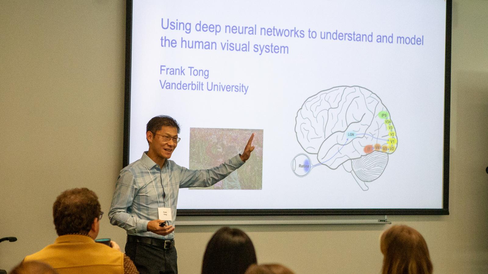
The 2023 CCBBI Research Day will feature a keynote address by Dr. Frank Tong, Centennial Professor of Psychology at Vanderbilt University; faculty presentations; and poster and short oral presentations by undergraduate and graduate students and research staff.
Keynote Address: Dr. Frank Tong

I grew up in Toronto Canada. I received my B.S. in Psychology at Queen's University in Kingston, Canada working with Barrie Frost, and my Ph.D. at Harvard University working with Ken Nakayama and Nancy Kanwisher. I worked with Steve Engel at UCLA for a year as a McDonnell-Pew post-doctoral fellow before starting my position as an assistant professor at Princeton University in 2000. My lab and I moved to Vanderbilt University in Fall 2004, where I am a Professor of Psychology. Awards and honors include the McDonnell-Pew Training Fellowship (1999), Robert K. Root Preceptorship, Princeton (2003), Scientific American Top 50 Award (2004), Young Investigator Award from the Cognitive Neuroscience Society (2006), Chancellor's Award for Research, Vanderbilt (2008), Young Investigator Award from the Vision
Sciences Society (2009), and the Troland Research Award in Psychology from the National Academy of Sciences. I have served as a board member of the Vision Sciences Society (2012-2016) and on the editorial committee of the Annual Review of Psychology (2013-2017).
Title: Using deep neural networks to understand and model the human visual system
Abstract: For this talk, I will begin by highlighting some of my favorite fMRI studies that emerged from my lab over the years, and discuss what they reveal about the computations and cognitive functions performed by the human visual system. Then I will discuss our ongoing efforts to determine whether deep neural networks (DNNs) can serve as useful models and testbeds to develop a better understanding of how the human visual system works. Although DNNs can match or outperform humans in many object recognition tasks, they tend to catastrophically fail in situations where humans do not, especially when faced with noisy, degraded, or ambiguous visual inputs. Such findings imply that the computations performed by DNNs do not adequately match those performed by the human brain. We will consider whether the brittleness of current DNN models is caused by flaws in their architectural design, imperfections in their learning protocols, or inadequacies in their training experiences. In particular, we will explore the hypothesis that visual blur may be a critical feature for conferring robustness to both biological and artificial visual systems.
Oral Presentations

Dr. Brandon Turner
Professor, Psychology
Distributed Neural Systems Support Flexible Attention Updating during Learning

Dr. Ivy Tso
Associate Professor, Psychiatry
Understanding altered eye gaze perception in schizophrenia using dynamic causal modeling

Dr. Scott Langenecker
Professor, Psychiatry
Predictive Models, Neural Phenotyping, and Disease and Lifespan Course of Mood Disorder

Dr. Baldwin Way
Associate Professor, Psychology
Does marijuana use impact adolescent neural development?
Behavioral and neural correlates of impaired scene perception following saccadic eye movements
Yong Min Choi, Tzu-Yao Chiu, and Julie D. Golomb
Department of Psychology, The Ohio State University
The visual input projected to the retina shifts drastically across saccadic eye movements. Although we are not aware of it, perception of simple visual stimuli presented around the time of a saccade is impaired (Burr et al., 1994). Meanwhile, whether/how the processing of complex visual scene images is impaired following saccades remains unclear. In the current study, we investigated scene perception following a saccade, through behavioral and fMRI experiments assisted by eye-tracking. In the behavioral experiment, subjects briefly viewed a scene image presented after different post-saccadic delays (5, 16, 50, 158, and 500 ms after saccade completion), and performed a 6-way categorization task on the scene content (beach, mountain, highway, etc). We found lower scene categorization accuracy in the 5 ms and 16 ms post-saccadic delay conditions compared to the baseline condition (500 ms), suggesting impaired scene perception shortly after the saccade which recovers back to the baseline within 50 ms post-saccade. In the fMRI study, subjects performed a 1-back task while making saccades and viewing scene images in the fMRI scanner. Short and long post-saccadic delay trials were selected post-hoc using eye-tracking data, and we used RSA-based decoding analyses to assess scene category information (urban vs nature scenes) in scene-selective brain areas, and low-level visual information (high vs low spatial frequency filtered images) in the early visual cortex. We found decreased scene category information in the posterior sub-region of the parahippocampal place area on short post-saccadic delay trials compared to long delay trials. Additionally, we found decreased spatial frequency information in the early visual cortex on short versus long post-saccadic delay trials. To summarize, the current study presents behavioral evidence of impaired scene perception following saccadic eye movements, supported by interrupted high-level scene category information AND low-level visual information of a scene image at different levels of visual hierarchies.
Keywords: Eye movement, Saccade, Scene Perception
Differences in the Relationship Between Structural Connectivity and Functional Activation Following Pediatric TBI
Florencia Ontiveros1,2, Whitney I. Mattson1, Kathryn Vannatta1,3, William A. Cunningham4, Elisabeth A. Wilde5, Keith O. Yeates6, and Kristen R. Hoskinson1,3
1Abigail Wexner Research Institute at Nationwide Children's Hospital, Columbus, OH,2The Ohio State University, College of Arts and Sciences, Neuroscience, Columbus, OH, 3The Ohio State University College of Medicine, Columbus, OH, 4University of Toronto, Toronto, ON, Canada, 5University of Utah, Salt Lake City, UT, 6University of Calgary, Calgary, AB, Canada
Introduction: White matter (WM) shear injury after traumatic brain injury (TBI) increases risk of WM degradation. TBI has been linked with spatial working memory (SWM) deficits, mainly subserved by the fronto-parietal network (FPN). WM connections within FPN functional hubs may be crucial. We explored associations among WM and FPN activation during SWM in children with moderate-to-severe TBI (msTBI), complicated-mild TBI (cmTBI), or orthopedic injury (OI).
Methods: Youth with msTBI (N=10, Mage=12.00), cmTBI (N=11, Mage=12.85), and OI (N=22, Mage=11.53) underwent MRI in a 3T Siemens scanner, including T1-weighted structural, 64-direction DTI, and task-based fMRI sequences. The SWM task involved characters living in linear space, information about an event at a location, and ranking arrival order. Freesurfer’s (v7.3.2) TRACULA performed automated probabilistic tractography on the corpus callosum (CC), cingulum bundles (CB), and bilateral anterior thalamic radiations (ATRs), longitudinal (i.e., ILF, MLF, SLF I-III), and uncinate fasciculi (UF). An FPN ROI (Neurosynth) was corrected (FDR<.01) and binarized. We extracted brain activation averages for high difficulty task trials relative to a control condition. We fitted a general linear model to WM measures at tract cross-sections, covarying for brain activation.
Results: Clusters reflect tract areas where association among WM and FPN activation differs by group. We found differences in factional anisotropy in the left dorsal CB and right SLF I, and mean diffusivity in parts of the CC, right ATR, bilateral CB, ILF, MLF, SLF I-II, left SLF III, and bilateral UF (ps<.05). The msTBI group had more variability in task performance than cmTBI and OI.
Discussion: We found group differences in WM microstructure of tracts related to FPN activation during SWM. Associations among structural connectivity and functional activation were different among groups, highlighting a possible deficit in SWM due to WM disruption post-TBI, improving our understanding of TBI-related brain chances subserve functional behavior.
Keywords: Traumatic brain injury, white matter, spatial working memory, frontoparietal network, structural connectivity, functional activation.
Object size and depth representations in human visual cortex
Mengxin Ran, Zitong Lu, and Julie Golomb
Department of Psychology, The Ohio State University
One of the key abilities in human object perception is having a stable representation of an object’s real-world size across various distances and perspectives. Previous research has indicated that neural responses in the ventral temporal cortex reflect object real-world size representations. However, the stimuli used in these prior studies confounded two related properties: perceived real-world size and real-world depth (distance). Moreover, the stimuli did not include naturalistic backgrounds, preventing us from exploring visual mechanisms in more ecological conditions. Bridging this gap, a recent study from our group conducted a model-based representational similarity analysis on EEG data from a large-scale dataset of subjects viewing natural images featuring objects of varying visual sizes and depths. The EEG study successfully disentangled neural representations of perceived object real-world size from perceived real-world depth and uncovered a distinct representational object processing timeline. To better understand object representations in human brains, our current study applies a similar analysis approach to fMRI data, aiming to explore how different parts of human visual cortex represent objects real-world size and depth information in natural images. Applying representational similarity analysis on the THINGS fMRI dataset, we isolated neural representations specific to real-world size, real-world depth, and visual size across human visual cortex. We found the most robust visual size representations in the Extrastriate Body Area, the most robust real-world depth representations in scene-selective regions such as the Parahippocampal Place Area and the Retrosplenial Cortex, and the most robust real-world size representations in face and object-selective regions, such as the Fusiform Face Area and the Lateral Occipital Complex. Our study delineates how various regions in human visual cortex are involved in processing different object size and depth features via an advanced computational approach, which offers an insightful understanding of the human brain processing of object information within naturalistic images.
Key words: Object representation; real-world size; visual size; real-world depth
Neural development of language and speech in infants and toddlers
Kelly J. Hiersche and Zeynep M. Saygin
Department of Psychology, The Ohio State University
Remarkably, children implicitly develop language skills very early in life. The “language-ready” brain theory suggests infant brains are prewired for language acquisition due to the asymmetries in anatomical regions associated with language processing (e.g., planum temporal). However, this hypothesis has not been directly tested, given that children are notoriously difficult to scan during these early years of language acquisition. We address these difficulties in both data acquisition and processing to examine the neural response to human speech and high-level linguistic properties and their associations with structural asymmetries both in anatomical regions traditionally associated with language processing, as well as subject-specific functional regions (fROIs). In infants (ages 2-35 months), we collected high-resolution structural data and multiple runs of a functional language localizer. We clean this data to only analyze timepoints with the lowest motion (less than 1°/mm between subsequent timepoints), and manually checked registration between functional and anatomical data. Following a GLM with sentences (containing high-level linguistic properties), reverse speech (fast temporal properties of language but devoid of syntax and semantics), and texturized speech (degraded auditory control) included as conditions (and including all timepoints with greater than .5 °/mm as nuisance regressors), we apply an individualized-subject approach, mirroring the rigor applied to adult neuroimaging studies, to create fROIs. We examine whether these fROIs differentiate high-level language and/or meaningless human speech in each subject. Then, we examine any relationships between speech or language selectivity and laterality with structural properties such as sulcal depth, cortical area, cortical thickness, and sulci/gyri curvature. To our knowledge, this is the first direct examination of the “language-ready” hypothesis in human infants using individualized functional activation and structural data and will provide important insights on the development of uniquely human functions.
Keywords: language, functional, structural, infant
Poster Presentation Abstracts
AKLO: A software protocol for site-specific neuromodulation delivery in brain-to-brain interface
Eric Zachariah1 and John LaRocco2
1Neurotech Club, 2Wexner Medical Center, The Ohio State University
Introduction: Brain-to-brain interface (BBI) involves a closed-loop system connecting a brain-computer interface (BCI) and computer-brain interface (CBI) among multiple users. As BBI requires low-latency site-specific neuromodulation delivery, a specialized bidirectional software protocol was developed.
Methods: Automated Kinetic Linking Optimization (AKLO) is an open-source framework for BBI built with existing, proven tools, including Python and Message Queue Telemetry Transport (MQTT). By combining AKLO with a stereotyped 10-20 International coordinate system, device-agnostic neuromodulation can be triggered, delivered, and confirmed. Data from an imagined speech electroencephalographic (EEG) BCI was used to validate a proof of concept by maximizing the information transfer rate (ITR) for the given paradigm. A simulated haptic stimulation was delivered to electrode positions C3 and C4.
Results: The imagined speech EEG BCI had a window size of 1 second, which constrained AKLO’s maximum ITR at 304.15 bits/min. When the update rate was adjusted to 100 ms, the ITR increased to 3041.5 bits/min. However, this value is still on par with a conventional keyboard or computer interface.
Discussion: Using archived data and simulating values from the literature, AKLO demonstrated it was a viable tool for BBI. The applications range from closed-loop medical devices to recreational use to non-verbal team communication. Future work will involve human trials and device optimization.
Keywords: Brain to brain interface, software protocol, computer brain interface, brain computer interface
Human EEG and artificial neural networks reveal disentangled representations of object real-world size in natural images
Zitong Lu and Julie D. Golomb
Department of Psychology, The Ohio State University
Human brains have the ability to accurately perceive and process the real-world size of objects, despite vast differences in distance and perspective, which is a remarkable feat of cognitive processing. While previous studies have delved into this phenomenon, our study uses an innovative approach to disentangle neural representations of object real-world size from visual size and perceived real-world depth in a way that was not previously possible. Our multi-modal approach incorporates computational modeling and the THINGS EEG2 dataset, which offers both high time-resolution human brain recordings and more ecologically valid naturalistic stimuli. Leveraging this state-of-the-art dataset, our EEG representational similarity results revealed a pure representation of object real-world size in human brains. We report a representational timeline of visual object processing: pixel-wise differences appeared first, then real-world depth and visual size, and finally, real-world size. Furthermore, representational comparisons with different artificial neural networks reveal real-world size as a stable and higher-level dimension in object space incorporating both visual and semantic information.
Keywords: real-world size, depth perception, RSA, artificial neural networks, object recognition
Comparative voltage gradient measurement of amplitude-modulated transcranial pulsed current stimulation in neural tissue phantoms
Taeyoon Eom1, Vania Gandhi1, Ekansh Seth1, and John LaRocco2
1Neurotech Club, 2Wexner Medical Center, The Ohio State University
Introduction: Amplitude-modulated transcranial pulsed current stimulation (am-tPCS) is a non-invasive electrical stimulation modality offering the potential to modulate along a region of interest (ROI). In order to optimize stimulation parameters, several brain tissue phantoms were prepared and compared. A cheap, expedient, and reliable phantom would capture much of the induced voltage gradient.
Methods: These included: agar gelatin mixed with salt at different weight ratios (1%, 5%, and 10% salt compositions), commercial ground beef, and phosphate buffered saline (PBS). The voltage gradients of a commercial am-tPCS device, operating at 5 V and 3.6 mA with 5-20 millisecond pulses, was evaluated in 3D across each phantom. The voltage gradients were compared for each.
Results: The observed voltage gradient was compared to manufacturer specified value of 3.8 mV/mm. An average voltage gradient of 1.21 mV/mm was observed, with the highest at 3.7 mV/mm in PBS, 2.9 mV/mm in ground beef, and 1.5 mV/mm in gel with 10% salt. While second highest in voltage gradient, the ground beef had the lowest cost ($10) and shortest preparation time (<1 hour).
Discussion: Ground beef was used in other studies as an electrical, optical, thermal, acoustic, and magnetic brain tissue phantom. Electrophysiological measurement with a ground beef phantom may be applicable for optimizing ROI-specific am-tPCS stimulation.
Keywords: brain model, electrical phantom, transcranial electrical stimulation, electrophysiology
Allocation of spatial attention in human visual cortex as a function of endogenous cue validity
William Narhi-Martinez, Yong-Min Choi, Blaire Dube, and Julie D. Golomb
Department of Psychology, The Ohio State University
Abstract: Certain areas of visual cortex are known to contain retinotopic maps of the visual field, and a number of neuroimaging studies have shown that covert attentional guidance will result in spatially-specific increases of neural activity within the cortical regions representing the attended locations. However, little research has been done to directly compare how attentional cues that carry differing levels of task-relevant information will impact cue-modulated neural activity. Here, we used fMRI with human participants to investigate how activity in area V4 would respond depending on if participants were cued with deterministic or probabilistic spatial information. Every trial began with a central arrow cue and subsequent pre-array delay, followed by a four-item memory array and subsequent post-array delay prior to the presentation of the memory probe for one of the memory array items. Critically, at the start of each run of trials, participants were informed that the arrow cues would indicate the to-be-probed item location with either 100% validity (deterministic spatial cue) or 70% validity (probabilistic spatial cue). Our results revealed greater activity in regions of V4 representing the cued location for a probabilistic relative to deterministic cue prior to the onset of the memory array, but greater cued-location activity for a deterministic relative to probabilistic cue following the onset of the memory array. These findings reveal how cue validity alone can drive the differential allocation of neural resources across cued and noncued locations. While information regarding where a target is most probable to appear can initially bias attention, this bias may be more likely to spread or wane over time. In contrast, information providing complete certainty regarding where the target will appear may bias attention for a longer period of time both in anticipation of, and following, the presentation of task-relevant stimuli.
Keywords: visual cognition, spatial attention, fMRI, endogenous cue, probabilistic
Altered Dynamic Functional Neural Connectivity (dFNC) in a Brain State Related to Default Mode (DMN) and Fronto-parietal (FPN) Network Connectivity in Acute Mindfulness and After an 8-week Mind-body Intervention in PTSD Patients
Garrett R. Hosterman, Lei Cao, Arushi Badola, Sophie A. Lazarus, and Anthony P. King
Department of Psychiatry and Behavioral Health, The Ohio State University
Introduction: Posttraumatic Stress Disorder (PTSD) is common and can be chronic and debilitating. Resting-state functional connectivity (rsFC) abnormalities have been observed in PTSD, in particular decreased rsFC connectivity between Default Mode Network (DMN) and Frontoparietal Network (FPN). Dynamic functional neural connectivity (dFNC) analysis identifies brain “states” based on time-course rsFC between various independent components (ICs), and how the brain switches between these states over time (“chonnectomics”). We tested dFNC changes in a pre-post design before and after an 8-week Mind-body intervention in PTSD patients.
Methods: Participants were N=41 persons from the community with a history of lifetime trauma with clinically significant symptoms of PTSD. We performed 3T fMRI before and after 8-weeks of a Mind-body intervention (Mindfulness or Progressive Relaxation Therapy) with two resting conditions: “Rest” – instructed to allow their mind to wander freely, and “Mindfulness” - focus their attention on thoughts, breath, and feelings. The GIFT toolbox was used to conduct joint ICA analysis, which identified components of neural connectivity within canonical networks (DMN, FPN, visual (VN), salience (SN), somatosensory (SSN) network, etc.). Sliding window analysis was run over the entire 8-minutes, with 32 second sliding windows. 8 brain states were identified, with mean dwell time calculated both before and after intervention.
Results: There was a significant decrease in mean dwell time in the “Mindfulness” vs “Rest” conditions in a brain state characterized by negative connectivity between the DMN and the FPN at intake (before Mindbody intervention). The same brain state decreased in the Rest condition from intake to post Mindbody intervention.
Discussion: dFNC revealed a single state in which mean dwell time during a standard rest from pre to post decreased to match dwell time during mindful rest. This could indicate a transition from more state mindfulness to trait mindfulness during rest.
Keywords: fMRI, rsFC, Trauma, PTSD, Randomized Control Trial, Frontoparietal Network, Default Mode Network, dFNC
Relationship between broad-scale functional connectivity and cognitive domains across individuals
Ajuna Mwesigye, Kelly J Hiersche, Zeynep Saygin, and David Osher
Department of Psychology, The Ohio State University
Abstract: Functional connectivity (FC) is related to functional activation; the location and extent of activation to a task can be predicted using the unique patterns of FC between the brain regions of an individual. Does this relationship extend to broad patterns of FC across individuals and the expected activation to a wide range of cognitive tasks? Do some cognitive domains show stronger associations with FC than others? Are some anatomical regions highly predictive of functional activation regardless of cognitive domain? To answer these questions, we obtained expected functional activation data for 27 cognitive domains using NeuroQuery (an online database that predicts activation maps based on semantic similarity among search queries and terms from a large set of previously published papers) and resting-state FC data from the Human Connectome Project. Across all individuals, we averaged the voxel to region (Glasser parcellation) FC matrix. This averaged connectivity data was the input to a ridge regression model. This model used the pattern of connectivity of each voxel in each region to all other regions to predict the expected functional activation within that region, for each cognitive domain. We compared the expected and model predicted activation of all regions for all domains; across all domains, connectivity explained a large proportion of the functional activation variance. Further, all regions showed strong association between connectivity and function, regardless of the cognitive domain, with greater variability across regions than was observed across domains. Overall, our results suggest strong associations between functional activation and connectivity across domains and regions.
Key words: Functional Connectivity, Functional activation, Cognitive domain
Increased Split-Half Reliability for Conditions vs Contrasts in fMRI analysis
Lingwen Ren, Athena L. Biggs, Stephanie Gorka, Luan Phan, and Jay C. Fournier
Department of Psychiatry and Behavioral Health, The Ohio State University
Introduction: Recent studies have questioned the reliability of metrics from task-based fMRI data. Establishing reliable fMRI measurements is critical for the clinically utility of neuroimaging. Here, we examined the internal consistency (i.e., split-half reliability) for an emotion regulation task. Additionally, we assessed how modeling decisions affected that reliability. We hypothesized there would be 1) higher reliability for task-based BOLD signals modelled within single conditions than condition contrasts, and 2) greater reliability in meta-analytic ROIs than study-specific ROIs.
Methods: Forty healthy participants completed an emotion regulation task that involved viewing neutral or negative images from the IAPS. Between each image, a fixation cross was shown. In the “Look Neutral” and the “Maintain Negative” conditions, participants naturally viewed images without attempting to change their emotional response. In the “Reappraise Negative” condition, participants were asked to change their thoughts about the image to alter their emotional response.
We calculated the adjusted split-half reliability for the condition and contrast signals specifically for Maintain and Reappraise conditions across 5 groups of ROIs: 8 from a meta-analysis (Morawetz et al., 2020) and 4 sample-specific (k>100, p<0.001): Maintain (16 ROIs), Reappraise (10 ROIs), Maintain-Fixation (7 ROIs), and Reappraise-Fixation (5 ROIs).
Results: Reliability for conditions alone (M rSB=0.49, range=[-0.341, 0.846]) was highest, followed by the Condition-Fixation contrasts (M rSB=0.36, range=[-0.816, 0.801]) and then the Condition-Look contrasts (M rSB=0.19, range=[-1.216, 0.616]), (F(2,153)=12.81, p<0.001). We observed no significant differences for meta-analytically-defined vs. study-specific ROIs.
Discussion: Reliability of the BOLD signal from task conditions alone showed higher internal consistency than contrasts. This suggests that future research using this task may be more likely to replicate if BOLD-signal extracted from conditions alone are prioritized over condition contrasts.
Keywords: BOLD reliability, internal consistency, emotion regulation
Similarity of Impression Towards the Characters is Related to Neural Synchrony While Watching Movie
Huanqing Wang and Dylan Wagner
Department of Psychology, The Ohio State University
Introduction: Although people are witnessing the same event, they always create different interpretations of it. This effect is amplified for social interaction events, as they are relatively ambiguous, and the way people perceive them can be influenced by many factors. However, which factors are related to or contributing to the individual differences has not yet been systematically investigated.
Methods:To answer this question, we recruited participants in a brain study, in which after they finished watching the first episode of the reality show The Mole, they reported their first impressions of the characters involved (pre-scanning). Then they went through a brain scanning in which they watched three independent videos from the show. After the brain scanning, they reported their post-scanning impressions of the characters.
Results: In this preliminary analysis, by using Inter-Subject Representational Similarity Analysis (IS-RSA), we found that the similarity of participants’ pre-scanning impressions of the characters is related to their brain temporal intersubject synchrony while watching the videos. We found that participants’ post-scanning impressions similarity is also related to their neural synchrony. Interestingly, we found that participants’ neural synchrony is more related to their similarity of cognitive impressions of the characters (i.e., the big-5 personality impression) rather than their similarity of affective impressions of the characters (i.e., how much the participants like/feel related to the characters).
Discussion: The results indicate that people who share similar impressions of the characters perceive the event more similarly. It is possible that while people are seeing social interaction events that happened to the characters, they have already put on an ‘impression glasses’ so that the way they interpret the events is biased by the impression they hold of the characters. Further analysis is needed to confirm this effect and to answer why cognitive impressions matter much more than affective impressions.
Sex differences in the prospective relationship between head hair cortisol levels and hippocampal volume
Kendra Wilson, Charles Ferris, Bethany Boettner, Ping Bai, Dylan Wagner, Christopher Browning, and
Baldwin Way
Department of Psychology, The Ohio State University
Background: In animal models, cortisol is a known driver of hippocampal volume and function, but less work has examined the cortisol-hippocampus pathway in humans (Kim, Pellman, & Kim, 2015). The adolescent brain may be more sensitive to glucocorticoids (Lee, Brandy, & Koenig, 2003) and animal studies suggest the hippocampus is more sensitive to glucocorticoids in males compared to females (Galea et al.,1997; Johnson et al., 2022), thus adolescent males may be particularly sensitive to the effects of glucocorticoids. We examine the longitudinal sex-specific effects of cortisol on hippocampal volume and predict that higher levels of head hair cortisol will predict reduced hippocampal volume measured years later, particularly in males.
Method: Head hair cortisol was measured over three months during wave 1(W1) of the Adolescent Health and Development in Context(AHDC) sample of youth ages 11 to 17(2014-2016). We acquired structural MRI data during wave 3(W3) of the AHDC study. 152 participants were included in the longitudinal analysis who provided cortisol at W1 and were scanned at W3 (57% female, Mage wave 1 = 13.0, Mage wave 3 = 16.1). We conducted moderation analyses in PROCESS (IV = W1 cortisol, DV= W3 hippocampal volume, Moderator = sex, Covariates= age, race, and caregiver’s highest level of education completed).
Results: Sex significantly moderated the effect of W1 cortisol on W3 hippocampal volume (p = .02). For males, W1 cortisol significantly and negatively predicted W3 hippocampal volume (p = .002, B = -.009). For females, there was no significant association (p = .99).
Conclusions: The results support animal models demonstrating sex-specific effects of cortisol on the hippocampus, and that cortisol levels in early adolescence can influence brain structure later in adolescence and early adulthood.
Key Words: Cortisol, Hippocampus, Stress, Sex Differences
Low Dose Ionizing Radiation Exposure in Repeat ED Encounters
Nick Basile1, Chad Donley2, Carl Peterson2, Timothy Barreiro2, and David Gemmel2
1Department of Biochemistry, The Ohio State University;
2Bon Secours Mercy Health St. Elizabeth’s Hospital, Youngstown, OH
Introduction: Low-dose ionizing radiation (IR) over a short period can accumulate, leading to an increased risk of DNA breakage or damage and other known health effects, such as oncologic risk. The purpose of this study was to measure the amount of IR exposure in frequent users of emergency services and to describe groups at increased risk.
Methodology: N= 714 patient records were abstracted from 404,000 encounters to the EDs between June 2021 and June 2023 in the Mercy Health Youngstown region. Total encounters by patients in 2 years were sampled and sorted by number of encounters. By observing the natural break in the number of encounters, a random sample of n=20 patients was taken if the number of encounters was 1-25 in 2 years at each encounter strata. For all patients with greater than 25 ED encounters in two years, 100 percent of patient records were reviewed (n=214). Amount and types of radiological imaging, demographics, and co-morbidities were also abstracted from the charts. Utilizing an ionizing radiation (IR) dosage table specific to the type of imaging and anatomical region, the amount of IR was accumulated for each patient in the 2 years in millisieverts (mSv). A comparison of two groups: > 100 mSv and < 100 mSv in the 2 years was undertaken, with 50 mSv considered significant for OSHA exposures in one year. Additionally, regression was undertaken to quantify the number of ED encounters required to reach OSHA thresholds for significant IR.
Results: Among this series of heavy ED users, the average age was 48 + 19 years, 60.6% were female, and 63.6 % were white. Medicaid or Medicaid managed care was the most prevalent guarantor. On average, the mean ED encounters over 2 years was 22 + 21 encounters in which patients obtained 11 + 12 CT scans. The average estimated IR exposure in this series over 2 years was 114 + 129 mSv. When ED encounters were modeled using linear regression for IR exposure at 100 mSv, at approximately n=17 ED encounters in two years, patients accumulated the at-risk dose threshold used by OSHA. Age was positively but weakly associated with IR (r=0.185, p < 0.001). While no difference in IR by race was noted, females exhibited higher IR exposure (p = 0.04). Total number of images, as expected, was highly correlated with IR (r=0.842, p < 0.001). Psych diagnoses were prevalent in the series and were statistically associated with increased IR exposure > 100 mSv: malingerer, noncompliance, depression/ bipolar, drug-seeking behavior, anxiety, chronic pain, fibromyalgia, alcohol abuse, and other psychiatric diagnoses (all p < 0.05 on Chi-Square).
Conclusions: Among very frequent ED users, especially poorer females with psych diagnoses, IR is problematic: half of the patients in this series exhibited mSv of exposure which exceeded those observed among Nagasaki survivors (in which median mSv was ~ 100). Extrapolating this data at the 100 mSv threshold over 2 years, IR exposure was not a problem for about 95.3% of encounters or 99.6% of people in the ED. Exposure represents a theoretical problem for about n=681 people (out of ~171,539) seen in the last 2 years in the EDs, about n=16,654 encounters (out of ~400k in the last 2 years) at 100 mSv. Recent data suggests a lower level of exposure at 50 mSv confers deleterious risk. Strategies to reduce IR among excessive ED users are needed.
Keywords: ionizing radiation, radiological imaging, emergency department
Feelings of Belongingness moderate the relationship between neural selectivity to cognitive demand and internalizing behaviors
Razan Mirghani, Elana Schettini, and Zeynep Saygin
Department of Psychology, The Ohio State University
Abstract: Previous literature shows that greater feelings of belongingness to someone’s culture and greater general cognitive control ability are both associated with fewer symptoms of psychopathology. Here, we test whether belongingness moderates the relationship between neural selectivity to cognitive load and internalizing symptoms. To do this, we use tabulated data downloaded from the two and three year follow up from the Adolescent Brain Cognitive Development Study, Release 5.0 (N = 2,176, 11-12 years). We use a task-dependent fMRI nBack task (2-back vs. 0-back contrast) to isolate neural selectivity to cognitive demand, the Child Behavioral Checklist (CBCL) to measure internalizing behaviors, and the Multi-Ethnic Identity Measure (Revised) to measure feelings of belongingness (i.e., how committed and accepted one feels towards their self-identified ethnic identity group). We use a multiple linear regression to examine the interaction between neural selectivity to cognitive demand and belongingness, while controlling for age; we tested this model separately for 18 bilateral frontal, opercular, cingulate, and parietal regions that are part of the domain-general multiple demand network. Each of our models replicate negative relationships for internalizing symptoms with belongingness, as well as with neural selectivity. We see that for the left and right caudal anterior cingulate there is virtually no relationship between neural selectivity to cognitive demand and the internalizing t score without belongingness included in the model. However, when belongingness is considered, the relationship between internalizing symptoms and neural selectivity is significantly different for those with low vs. high feelings of belongingness. For the left superior frontal gyrus, left pars opercularis, supramarginal gyrus, and the insula we see a negative relationship between neural selectivity to cognitive demand and internalizing scores when belongingness is not accounted for. Adding belongingness to the model strengthens the negative relationship for the group with low feelings of belongingness, and there is virtually no relationship for those with high feelings of belongingness. These results may suggest that greater feelings of belongingness are a protective factor against internalizing symptoms in children, regardless of their neural selectivity to cognitive demand. These findings may inform future research examining malleable factors that could attenuate risk to psychopathology in children.
Key Words: Internalizing behaviors, cognitive demand, nBack task, executive network, belongingness
White Matter Connectivity of FFA and VWFA to Higher-Level Cortex Involved in Language and Theory of Mind
Leah DiRubio, Jin Li, and Zeynep Saygin
Department of Psychology, The Ohio State University
Abstract: The Fusiform Face Area (FFA) and Visual Word Form Area (VWFA) are two regions of high level visual cortex selective to faces and visual words, respectively. The FFA and VWFA are within close anatomical proximity but are functionally distinct. Previous work has hypothesized that structural connectivity to language regions may play a role in the localization of the VWFA. Little work has explored the structural connectivity of the FFA beyond local connections to other face-processing regions. The primary goal of this study is to elucidate the FFA and VWFA’s patterns of structural connectivity to higher level regions involved in the related functions of language and theory of mind (ToM), and to compare their patterns of connectivity. Four functional tasks were used to localize the VWFA and FFA, as well as regions involved in language and theory of mind in 35 neurotypical adults. Then, structural connectivity, measured with diffusion weighted imaging, was estimated between these subject-specific fROIs using probabilistic tractography. Preliminary results showed that the lVWFA showed privileged connectivity (as compared to lFFA) to the left temporal language regions. Interestingly, TOM regions showed similar connectivity with both VWFA and FFA in both hemispheres, albeit with higher connectivity to both visual regions on the right than left. We discuss how the functional specialization of high-level visual cortex may be biased by their connectivity with higher-order cortices.
Keywords: Fusiform Face Area, Visual Word Form Area, Language, Theory of Mind, Structural Connectivity
Aberrant Functional Connectivity (rsFC) Between Subgenual ACC and Default Mode Network in Adults with History of Childhood Maltreatment
Arushi Badola, Subhashini Madhavan, Garrett Hosterman, and Anthony King
Department of Psychiatry and Behavioral Health, The Ohio State University
Introduction: Childhood maltreatment (CM) involves exposure to physical, sexual, and emotional abuse or neglect. CM increases one’s risk of developing negative physical and mental health outcomes. Previous studies have linked CM in Major Depressive Disorder (MDD) patients to increased functional connectivity (FC) between the default mode network (DMN) and the salience network (SAN). This study examined the resting-state functional connectivity (rsFC) patterns specific to CM in the DMN, SAN and limbic networks in adults to identify the neural correlates of CM.
Methods: N= 56 adults (20 with CM) with trauma exposure and symptoms of PTSD were recruited. Functional MRI scans were collected while participants were instructed to focus on a fixation “plus sign” and let their minds wander freely. Seed-based subject level rsFC T-maps (first level) were generated using CONN toolbox and entered into second-level random effects model in SPM12 to test the main effects of CM. The rsFC between individuals with and without CM were compared by examining the sgACC, ventromedial prefrontal cortex (vmPFC) and rostral anterior cingulate cortex (rACC).
Results: Participants with CM showed greater rsFC between the sgACC and the vmPFC ([2,52,-2]; kE=87; Z=4.15; whole-brain cluster pFDR = 0.013), increased connectivity between the rostral ACC and the vmPFC ([-4,28,-8]; kE=129; Z=4.28; whole-brain cluster pFDR = 0.005), and increased connectivity within the sgACC ([8,38,-20]; kE=100; Z=3.99; whole-brain cluster pFDR =0.006) compared to participants without CM.
Discussion: The results revealed aberrantly increased rsFC between the sgACC and nodes of DMN in persons with CM, consistent with previous reports of increased sgACC-DMN rsFC related to depressive rumination. Future studies may use these findings to study other factors related to CM and improve our understanding of the neurobiology of CM.
Keywords: Childhood Maltreatment, Default Mode Network, Salience Network, Resting-State Functional Connectivity, Subgenual ACC
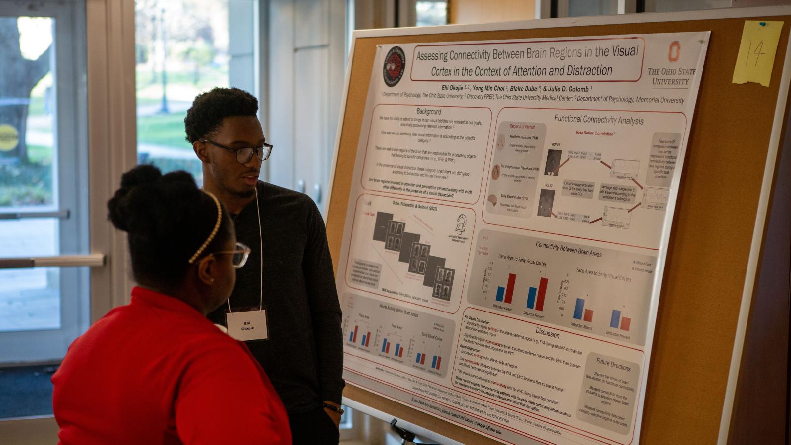
Assessing Connectivity Between Brain Regions in the Visual Cortex in the Context of Attention and Distraction
Ehi Okojie, Yong Min Choi, Blaire Dube, and Julie Golomb
Department of Psychology, The Ohio State University
Abstract: The brain has ways of processing information to prioritize input relevant to our goals, filtering out less relevant input. There are well-known brain regions responsible for processing objects belonging to specific categories, such as faces and scenes. These brain areas can act as attentional filters biased toward processing information relevant to our goals. A question we have is: What happens to these category-tuned filters when we get distracted? A recent fMRI study by Dube et al., 2022, investigated the effects that visual distraction has on the category-tuned filters in the ventral visual cortex (Fusiform Face Area and Parahippocampal Place Area) and discovered a novel consequence of distraction on these filters which regulate categoryspecific object processing. Participants in this study viewed hybrid face/house images and were told to attend to either faces or houses. They found that the presence of a salient distractor disrupts these filters, and our brain incidentally processes the goal-irrelevant category more than during distractorabsent trials. The current study expands on this finding, asking what brain areas may be modulating this filter disruption, to learn more about the neural pathways implicated in this filter disruption. We conducted univariate and functional connectivity analyses among particular brain regions during distractor-present versus distractor-absent trials. We compared connectivity between the task-relevant category-tuned area (FFA or PPA) and the early visual cortex (EVC). Results show that during distractor-absent attend-face trials, the FFA shows significantly higher connectivity with the EVC than during distractor-absent attend-house trials. This connectivity difference is nonsignificant during distractor-present trials. In attend-face blocks, the PPA shows numerically higher connectivity with the EVC during distractor-present trials than distractor-present trials. These results suggest that connectivity patterns with the early visual cortex may inform us about the mechanism underlying category-selective attentional filter disruption.
Keywords: Attention, distraction, filter disruption, visual perception, functional connectivity, fMRI
ADHD Symptom Severity and Neural Reward Processing in Children
Divya Gopal, Elana Schettini, and Zeynep Saygin
Department of Psychology, The Ohio State University
Abstract: Prior studies show differences in neural reward processing between children with and without attention-deficit hyperactivity disorder (ADHD). In this study, we investigate the relationship between ADHD symptoms and brain activity while children are anticipating reward within several subcortical and frontal cortical brain regions previously implicated in reward processing, including the bilateral striatum (caudate, putamen, nucleus accumbens), amygdala, insula, and anterior cingulate cortex (ACC). Previous research shows that when anticipating reward, individuals with ADHD show hyperactivation in cortical regions, such as the anterior cingulate cortex, and hypo-activation in striatal regions. We use the Cash Choice Task (CCT) and a task-dependent fMRI Monetary Incentive Delay (MID) task to explain variability in the sum score of ADHD inattentive and hyperactive symptoms, separately, from the Kiddie Schedule for Affective Disorders and Schizophrenia (K-SADS). Tabulated data was downloaded from the baseline Adolescent Brain Cognitive Development (ABCD) study Release 5.0 (N = 4,785, 9-10 years). We included children who had completed the CCT, MID-task, and KSADS; children were excluded if they were diagnosed with a bipolar, a psychotic, or autism spectrum disorder, or had a history of traumatic brain injury with any loss of consciousness. We used poisson regressions to explain variance of the symptom counts from CCT responses and MID regional activation (anticipation of Reward vs. Neutral), separately. We saw no significant relationships between CCT and ADHD symptom severity. For brain reactivity to reward, inattentive symptoms were negatively related to activation in the left putamen and positively related to activation in the bilateral amygdala and rostral ACC. Hyperactive symptoms were also negatively related to activation in the left putamen and positively related to activation in the bilateral amygdala and right accumbens area. These results demonstrate a dimensional relationship between severity of inattention and hyperactivity and neural reactivity to anticipation of reward.
Keywords: ADHD, Reward Processing, Ventral Striatum, Inattention, Hyperactivity, Anticipation of Reward
Research Day Committees
Uyen Tram, CCBBI Manager
Adam Gorka, Research Assistant Professor
James Teng, Graduate Research Assistant
Xiangrui Li, CCBBI Assistant Director
Elizabeth Sponseller, MRI Technologist
Ruchika Prakash, CCBBI Director
Kelly Hiersche
Eunjee Ko
Jenny Lin
Dan Zhu
Jay Fournier, Psychiatry and Behavioral Health
Jasmeet Hayes, Psychology
Ruchika Prakash, Psychology
Zeynep Saygin, Psychology
Julie Golomb, Psychology
Adam Gorka, Psychology
Kristen Hoskinson, Nationwide Children’s Hospital
Andy Leber, Psychology
David Osher, Psychology
Madhura Phansikar, Psychology
Dylan Wagner, Psychology
Baldwin Way, Psychology


