Equipment
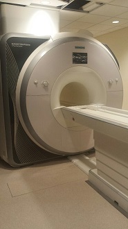
Siemens Prisma 3 Tesla MRI scanner with integrated Total Imaging Matrix (TIM) system
Siemens TIM is a high throughput imaging technology that allows for faster scanning in optimal spatial and temporal resolutions. It improves statistical power of MRI and functional brain mapping greatly. At CCBBI, Prisma 3T delivers high quality fMRI series with increased signal-to-noise ratio and reduced susceptibility artifacts. The outstanding gradient performance (80 mT/m at 200 T/m/s simultaneous on all three axes) leads to exceptionally high-quality diffusion-weighted imaging (DWI) and excellent robustness overall. The parallel transmit (pTX) technology TimTX TrueShape enables selective excitation of specific body areas through independent channels. Multi-band technology further accelerates functional and diffusion image acquisition. The 60 cm-diameter bore, 100% duty-cycle capability of the gradients, and the transmit/receive head RF coils make it ideal for human neuroimaging applications within current FDA peripheral nerve stimulation guidelines.
Key Attributes of the System
High field strength
High-field 3 Tesla magnets provide the optimal quality and susceptibility balance for human imaging. The increased soft-tissue contrast at 3T in combination with fast and ultra-fast pulses (i.e. in the order of single milliseconds) reveals fine biological structure. Scientific and clinical inquires in the neurosciences are particularly complex due to the fast nature of neuronal communication, and its metabolic surrogates are measurable only in specific magnetic conditions. Increased spatiotemporal resolution is critical for quality neuroimaging. Our MRI scanner provides accurate functional images of the human brain due to increased blood-oxygen level dependent (BOLD) contrast in our setting, favoring high functional contrast-to-noise ratio and faster scans.
Stable and proven design
Prisma is the latest of the Siemens Magnetom systems that are widely used in major research settings. Siemens has a track record of stability and reliability worldwide.
Comprehensive and versatile scan options
- Gradient-echo and spin-echo sequences (multi-planar or 3D) provide a wide range of tissue contrast (T1-, PD- and T2-weighted contrast) for structural imaging. With these sequences, MRI parameters (such as T1, T2, and proton density) can be quantified accurately. Sophisticated intra tissue properties can be inferred from special T1-rho and T2* sequences as well as quantitative iron concentration measurements and other study-customized pulse options.
- Spin-echo sequences (multi-echo/multi-plane, inversion recovery and derivatives). These are particularly useful for automated tissue segmentation. They are ideally suited to the creation of a flat cortical map of each individual subject.
- Fast spin-echo sequences (2D and 3D), which combine the advantages of spin echo techniques with higher resolution and faster acquisition times. These sequences allow us to acquire very high-resolution images (1024x1024); 3D datasets with cubic voxels, datasets with very thin slices through a combination of multiple interleaved 3D slabs, and also to perform flow compensation and magnetization transfer imaging.
- Diffusion-weighted MRI (DWI) can recover more anatomic sub-structure that cannot be resolved by conventional structural MRI methods. Diffusion tensor imaging (DTI) tractography allows the visualization of multiple white matter tracts based on diffusion tensor imaging data, and capture better visualizations of brain connectivity. DTI tractography in our system delivers up to 256 diffusion directions. We now have the ability to acquire high-resolution, ultra-fast, and highly reproducible 64 and 97 directions DTIs in multi-band.
- Echo planar imaging (EPI) with a full database of pulse sequences for very rapid acquisitions is ideally suited for functional MRI. The ultra-fast image reconstruction and real-time rendering packages run on the operator’s console. Our fMRI package employs Siemens TIM technology to orchestrate full and partial k-space filling and is designed for excellent Parallel Imaging in all directions. It greatly improves spatial-temporal resolution, signal-to-noise ratio and minimizes image distortion in functional MR imaging.
- 3D PACE prospective motion correction is used for reducing the influence of head movement on functional MRI and improves data robustness and reproducibility.
- The multi-band technology accelerates image acquisition in all contrast modalities, significantly enhancing the quality of data acquisition in neuroimaging studies.