Research Day 2024 Program Book

Keynote Address: Dr. Natasha Rajah

Dr. Natasha Rajah received her Ph.D. from the University of Toronto, St. George Campus; and did her postdoctoral training at U.C. Berkeley. She was a Professor at McGill University from 2005 to 2023 before joining the Department of Psychology at Toronto Metropolitan University (formerly Ryerson) in Fall 2023 as a tenured Full Professor and Tier 1 Canada Research Chair in Sex, Gender, and Diversity in Brain Health, Memory and Aging. Dr. Rajah uses multidisciplinary methods, including MRI, to investigate how sex, gender, and social determinants of health contribute to individual differences in brain and cognitive function in aging. She is an Editor-in-Chief for Aging, Neuropsychology and Cognition, Senior Editor for Brain Research and serves as Vice-Chair of the CIHR’s Institute of Aging Advisory Board; and is the EDI Lead for the Canadian Consortium on Neurodegeneration in Aging (CCNA) in Phase III.
Title: The effect of biological sex and menopause status on episodic memory and related brain function at midlife: The BHAMM Study
Abstract: Our ability to encode, store and retrieve past experiences in rich contextual details (episodic memory) declines with age and can be an early sign of Alzheimer’s disease (AD) – a disease that is more prevalent in females than males. Recent evidence also suggests that midlife is a critical period in adulthood when episodic memory decline and changes in brain regions affected by AD arise. Midlife is also the time that females with ovaries experience spontaneous menopause. Yet, little is known about how biological sex, menopause, and age affect episodic memory and related brain systems at midlife; and how this may differ in adult with risk factors for AD. In this talk I will present results from the ongoing Brain Health at Midlife and Menopause (BHAMM) study which aims to fill this knowledge gap and determine what factors support and/or hinder memory and brain function in males and females at midlife. Our initial results suggest that middle-aged females, but not males, exhibited age-related episodic memory decline, and that this effect was observed at post-menopause. Task-based fMRI analysis shows that memory decline at post-menopause was associated with altered activity in parahippocampal, posterior parietal and occipital cortices. Together these studies highlight the importance of considering biological sex and endocrine aging in the cognitive neuroscience of aging and dementia prevention.
View the talk below.
Featured Alumni Presentations

Dr. Paul Scotti is Head of Neuroimaging & AI at Stability AI and a visiting scientist at Princeton University, where he did his postdoc with Dr. Ken Norman. Paul leads the MedARC Neuroimaging & AI Lab which adopts an open lab concept where the research team is almost entirely composed of volunteers working together on open-source neuroAI projects. Paul did his Ph.D at The Ohio State University co-advised by Dr. Julie Golomb and Dr. Andy Leber, where he worked on projects including attraction/repulsion dynamics surrounding visual mnemonic representations and novel improvements to the inverted encoding model technique used in neuroimaging.
Title: Reconstructing seen images from fMRI brain activity
Abstract: I will discuss how recent AI advances have enabled high-quality reconstructions of seen images from solely fMRI brain activity. Specifically, we leveraged large-scale fMRI data to align brain activity to the pre-trained embedding space of CLIP via diffusion models and contrastive learning. Subsequently we developed a simple approach to achieve high-quality reconstruction results with just 1 hour of fMRI training data by aligning multiple participants to a shared latent space.

Charlie Ferris is a postdoctoral fellow at McGill University where he researches neural mechanisms supporting memory in humans. Charlie earned his Ph.D. at Emory University in Georgia working with Dr. Stephan Hamann, and then completed his first post-doc at The Ohio State University under the supervision of Dr. Baldwin Way. By combining fMRI, brain stimulation, and behavioral approaches, Charlie tests how brain networks dynamically process different types of memory content. As Charlie’s current work investigates episodic memory formation, consolidation, and retrieval using naturalistic stimuli in humans, he recommends you read this bio carefully as there may be a quiz on this information during his talk.
Title: Hippocampal-cortical networks predict conceptual versus perceptually guided narrative memory
Abstract: If the same basic story is told with a focus on different details, is the memory representation for the core elements of the story the same? Or does the underlying memory trace shift to match the surrounding details? Current theories of event memory propose distinct connections between the hippocampus and neocortical regions to support processing different types of content in memory. Additionally, it has been established that hippocampal connectivity supports integrating disparate content into unified event memories. This suggests that changing the way that an event is described could change the underlying neural representation of integrated event memories. We tested this proposition by developing event narratives that described the same core story with identical central story details (e.g., grocery shopping), but described with additional descriptive details related to the story that were conceptual or perceptual in nature. Using fMRI, we established hippocampal connectivity patterns as a group of human participants encoded these narratives, and then related these patterns to later memory for the narrative details. We found distinct patterns of hippocampal-cortical connectivity during encoding of the identical central event details when they were embedded between conceptual vs. perceptual details.

Dr. Stephanie Aghamoosa is a clinical neuropsychologist and Assistant Professor at the Medical University of South Carolina with expertise in using cognitive assessments and fMRI to study aging and Alzheimer's disease. She obtained her PhD at Ohio State in 2020 under the mentorship of Dr. Ruchika Prakash, conducting research on characterizing age-related neurocognitive changes and testing behavioral interventions to enhance cognitive and emotional function in older adults. She then completed a postdoctoral fellowship specializing in the clinical neuropsychology and cognitive neuroscience of early Alzheimer’s disease at the Medical University of South Carolina, where she is now a tenure-track faculty member. Her ongoing research is focused on developing non-pharmacological interventions to slow the clinical progression of Alzheimer’s disease. She is conducting technology-leveraged clinical trials of non-invasive brain stimulation and cognitive training designed to maximize neuroplasticity, using fMRI and digital health technology to sensitively measure intervention effects.
Title: Leveraging Functional Connectivity to Develop Effective Interventions for Early Alzheimer’s Disease
Abstract: Alzheimer’s disease (AD) is characterized by progressive cognitive impairments that are accompanied by widespread alterations in brain structure and function. The early stages of AD represent a critical window of opportunity for disease detection and intervention, but changes in cognition and brain function during this period are subtle and difficult to detect. However, advanced fMRI analysis techniques offer a promising avenue for sensitively characterizing and tracking early AD-related network alterations. In this talk, I will review my work using functional connectivity to reveal brain network disruptions that relate to cognitive deficits in preclinical AD. I will then discuss our clinical trials of non-invasive brain stimulation for improving cognition in mild cognitive impairment due to AD that integrate functional connectivity as an outcome measure to capture treatment effects, investigate neural mechanisms, and guide intervention development. I will discuss how recent AI advances have enabled high-quality reconstructions of seen images from solely fMRI brain activity. Specifically, we leveraged large-scale fMRI data to align brain activity to the pre-trained embedding space of CLIP via diffusion models and contrastive learning. Subsequently we developed a simple approach to achieve high-quality reconstruction results with just 1 hour of fMRI training data by aligning multiple participants to a shared latent space.
Watch the videos below.
Dr. Paul Scotti
Dr. Charlie Ferris
Dr. Stephanie Aghamoosa
Student Oral Presentations
Oral Presentation Abstract 1
Examining Developmental Variability in Functional Selectivity and Spatial Location of High-Level Visual Regions
Kelly J. Hiersche, Anna L. Quatrale, Zeynep M. Saygin
Department of Psychology, The Ohio State University
Introduction: The brain is a patchwork of regions, each devoted to different mental functions. Some of the most robust and replicable regions include high-level visual areas within ventral temporal cortex (VTC), such as the word-selective visual word form area (VWFA), face-selective fusiform face area (FFA), and object-selective posterior fusiform sulcus (PFS). Face and object regions show category selectivity very early in development, whereas word regions develop after literacy. These regions show high inter-subject variability in adults, but how does the variability of the selectivity and location of these regions change with age?
Methods: We collected structural data, and two runs of a high-level visual localizer from 112 children (including repeated timepoints) and 64 adults. From this sample, children were grouped by age and motion matched across groups (24 per group, ages 3-6, 6-9, and 9-12 years), and to an adult reference group (N=17). We defined subject specific functional regions of interest (for VWFA, FFA, and PFS), used independent data to calculate selectivity to the preferred category, and determined the location of the fROI along the VTC based on each subject's parcellated brain. Using the adults as a reference, we calculated the coefficient of variation, a measure of variability, for the selectivity and location of each fROI for each child and examined age related changes.
Results: We found variability of selectivity in the LH VWFA increases with age, whereas variability of selectivity decreases in bilateral FFA. Further, location variability of the LH VWFA increases with age, and LH VWFA shows a medial shift across development. This may be due to brain growth, as the youngest children also show significantly greater cortical thickness in most lateral sections of the VTC; however, cortical thickness of an fROI did not explain its location, selectivity, or the variability in either of those measures.
Discussion: Increases in variability of the LH VWFA suggest that word specialization diverges over development and as literacy develops, whereas variability of face specialization converges towards adults over development. While cortical thickness is likely important for large-scale organization and development, it does not account for the variability of fROI location or responses. We next plan to examine if functional connectivity to functional-defined networks (e.g., language) may explain these variability differences across ages and individuals.
Keywords: Development, functional regions of interest, variability
Oral Presentation Abstract 2
How do we store Kanizsa illusions in visual working memory?
Jenny Lin, Lisa M. Heisterberg, Mackenzie J. Siesel, and Andrew B. Leber
Department of Psychology, The Ohio State University
Introduction: Visual working memory is limited in capacity, but we can use various strategies to improve performa2 items that form an illusory shape (Kanizsa illusion) versus items that are randomly oriented (Allon et al., 2019). However, the mechanism behind this benefit is still unclear. Some have proposed that Kanizsa illusions improve performance by reducing the number of items stored in working memory. Previous studies tested this idea using neuroimaging methods to indirectly measure working memory load during task performance. Specifically, researchers have measured the contralateral delay activity (CDA), an event-related potential component shown to track working memory load. Thus far, there is mixed evidence that Kanizsa illusions reduce storage demands (Heisterberg, 2021; McCollough, 2011; Ping, 2020; see also Diaz et al., 2021). Here we further explore the role of encoding time in the Kanizsa benefit. Specifically, we examine whether participants require longer encoding time to integrate the illusory shape and reduce working memory load.
Methods: We had participants perform an orientation change detection task with EEG recording (N = 39; this preregistered study is in progress with a total planned sample of N = 80). On each trial, an arrow cued participants to attend to the left or right side of the screen. Next, participants saw memory stimuli consisting of either one “pacman” stimulus (single), three randomly oriented stimuli that form a triangle-like configuration (proximity), or three stimuli that form an illusory triangle (Kanizsa). Following the retention delay, participants responded whether the orientation of the test pacman is same or different as the memorized item. Critically, we manipulated encoding time of the memory display (short vs. long) between subjects. The encoding time was 200 ms in the short encoding group and 1000 ms in the long encoding group.
Results: Preliminary results show a Kanizsa benefit for both short and long encoding groups: memory accuracy was higher for the Kanizsa versus proximity condition. There was a greater CDA amplitude for proximity versus single condition for both the short and long encoding group. This replicates the finding that CDA is sensitive to working memory load. Critically, we found a lower CDA amplitude for the Kanizsa compared to the proximity condition in the long encoding group.However, we did not find a statistically reliable difference between these two conditions in the short encoding group.
Discussion: These findings might suggest that Kanizsa illusions reduce storage demands when there is sufficient encoding time for participants to use chunking strategies.
Keywords: visual working memory, strategy use, contralateral delay activity
Oral Presentation Abstract 3
Investigating Cerebellar Grey-Matter Volume Differences Across Brain Regions in Schizophrenia Versus Bipolar Disorder
Jayden Morales, Jessica Turner, Mahmoud Rashidi
The Ohio State University
Introduction. Schizophrenia (SZ) and bipolar disorder (BP) are debilitating mental illnesses that pose significant challenges for affected individuals. Symptoms of these conditions are associated with shared abnormalities in brain structure. Previous studies using neuroimaging techniques have revealed patterns of volume reduction in cortical regions, relative to healthy controls. This study examined volume differences across cerebellar gray- matter regions to identify distinctions between patients with bipolar disorder, patients with schizophrenia, and healthy controls,
Methods. Participants were N=289 adults-subjects, including 52 patients with bipolar disorder, 89 patients with schizophrenia, 34 first-degree relatives of a patient with bipolar disorder, 56 first-degree relatives of a patient with bipolar disorder, and 51 healthy controls. All patient data was collected at the University of Minnesota. The automatic cerebellum anatomical parcellation using U-Net with locally constrained optimization (ACAPULCO) algorithm was used to perform a segmentation of the cerebellar lobules across 28 brain regions. Volumes associated with segmented cerebellar lobules were analyzed using linear regression models to measure individual differences between participant groups. Covariates including age, gender, and intracranial volume (ICV) were included in the models to account for the variability they bring forth.
Results. Participants with Schizophrenia show reduced total cerebellar gray-matter volume relative to controls (p = 0.038). This effect no longer remained significant when gender, age, and ICV were uncontrolled. When SZ patients were compared to controls, the largest effects were found in the left Crus I, right Crus II, right VIIB, and left VIIB, with respect to order. All of these brain regions showed statistically significant reductions in volume. When BP patients were compared to controls, the largest effects were found in the left Crus I, left VIIA, left VIIB, and left V, with respect to order. None of these brain regions showed statistically significant reductions in volume. When SZ patients were compared to BP patients, the largest effects were found for total cerebellar volume (TCV), left Crus II, right Crus II, and right VIIB, with respect to order. All of these regions showed statistically significant reductions in volume. Interestingly, there were four brain regions where BP patients showed a larger magnitudinal effect in volume compared to SZ patients, relative to controls. These regions included the Vermis X, right VIIIB, left VIIIB, and left Crus II.
Discussion. Our results suggest that individuals with schizophrenia have notable volumetric reduction patterns across cerebellar regions, relative to both controls and patients with bipolar disorder. This indicates that there is in-fact a difference between patients with schizophrenia and patients with bipolar disorder in-terms of their cerebellar volume. None of the effects demonstrated by the BP group showed statistical significance, further analysis of this group in the future is needed. Furthermore, similar volume reduction patterns are found between patients with schizophrenia and patients with bipolar disorder relative to controls in the left Crus I, left VIIIA, left VIIB, left V, Vermis IX, Vermis VI, and a few other brain regions. While these volumetric reduction patterns are similar, they are significantly more apparent in the SZ group compared to the BP group.
Keywords. schizophrenia, bipolar disorder, cerebellar volumes, cerebellum, ACAPULCO, brain lobules
Oral Presentation Abstract 4
Connectomic reorganization following an 8-week mindfulness-based intervention in older adults
James Teng1, Anita Shankar1, Megan E. Fisher1, Nathan R. McPherson1, Madhura Phansikar1, Rebecca Andridge1,2, and Ruchika S. Prakash1.
1Department of Psychology, The Ohio State University, 2Division of Biostatistics, The Ohio State University
Introduction. Aging is associated with decreases in a multitude of cognitive and neural domains (Fortenbaugh et al., 2015; Koen and Rugg, 2019). To mitigate these age-related declines, behavioral interventions such as mindfulness have shown initial promise that must be investigated with methodological rigor, as the neural reorganization that accompanies these improvements remain poorly understood (Cotier et al. 2017). Entropy, a measure of network uniformity and specialization (Wang et al., 2014), can be analogous to neural dedifferentiation, and recent work has demonstrated increases in entropy with advancing age, which moderated cognitive declines (Shankar et. al, 2024). The current study examined within-sample entropy changes over a one-year period in older adults (Prakash et al., 2022) and examine the short-term (8 weeks) and long-term (one year) ability of mindfulness training to alter trajectories of network reorganization, at nodal, network, and whole-brain levels. Especially pertinent are the Default Mode network (DMN; Kral et al., 2019) and its posterior cingulate cortex (PCC) nodes, and the Frontoparietal Network (FPN) with its Inferior Frontal Gyrus (IFG) nodes implicated in mindfulness-related improvements in cognition (Taren et al., 2017).
Methods. 107 Older adults (aged 65 – 85) participated in the 8-week HealthyAgers randomized clinical trial with two intervention arms: Mindfulness-based stress reduction (N = 52) and lifestyle education (N = 55). Functional MRI (fMRI) data was collected at three timepoints (pre-intervention, post-intervention, and at 12-month follow-up) during rest and during a gradual-onset continuous performance task (gradCPT, Rosenberg et al., 2013). Functional connectivity was calculated from time-series Pearson's correlations for each node-pair of the Shen 268 atlas. From these, edge time series was derived by multiplying element-wise z-scored parcel time series for each node-pair in the connectivity matrix (Faskowitz et al., 2020). Next, k-means clustering was performed on the edge time series to assign a community label for each edge, before the distribution of edge community participation is calculated. Entropy is thus calculated as:
Where hi is the entropy of the ith node and pic is the fraction of the connections in the ith node assigned to edge community c. Average entropy scores were calculated for the whole brain, the DMN and FPN, and nodes of the PCC and IFG.
Results. Task fMRI showed no significant short- or long-term decreases in entropy across the whole-brain, nor within networks and nodes. However, across all participants, entropy was significantly reduced in resting state fMRI at a whole-brain level (F(2,144.4)=5.52, p=.0049), as well as within the DMN (F(2,147.9)=4.40, p=.0139), and FPN (F(2,154.1)=5.52, p=.0035). Nodal entropy did not significantly decrease, nor were there differential effects of the intervention on entropy.
Discussion. While entropy was significantly improved at both a whole-brain and network level during resting state, improvements in task performance were only seen at a nodal level. This suggests that age-related resting entropic changes may be enhanced with standardized 8-week mind-body interventions, though the granularity of these effects suggests the benefits may not be uniform. Nevertheless, our results offer potent intervention candidates for addressing the deleterious effects of aging on cognitive function.
Keywords: Network Neuroscience, Neural Reorganisation, fMRI, Entropy, Aging
Watch the video below.
Poster Presentations
Poster Presentation Abstract 1
Links among subcortical brain volume and social and emotional outcome in pediatric congenital heart disease
Declan E. Alford1, Florencia Ontiveros1, Aaron McAllister, M.D.2,3, May Ling Mah, M.D.2,3, Kathryn Vannatta, Ph.D.1,3, Kristen R. Hoskinson, Ph.D.1,3
1Abigail Wexner Research Institute at Nationwide Children’s Hospital, 2Nationwide Children’s Hospital, 3The Ohio State University College of Medicine
Introduction: Across specific diagnoses, pediatric congenital heart diseases (CHD) can disrupt the oxygenation of blood circulating in the central nervous system, which could impact subcortical brain areas, including through volume reduction. This may contribute to comorbidities faced by CHD patients, including neurodevelopmental disruptions to behavioral and emotional function. Examining links among neural and behavioral sequelae of childhood CHD is essential, as a growing population survives into adolescence and into adulthood. This project explores the relationship among various subcortical volumes and behavioral implications in youth with CHD relative to healthy children (HC).
Methods: This project included children with CHD (n=23, Mage=11.71yr, Male=13), and HC (n=14, Mage=11.47yr, Male=12). Participants completed functional and structural MRI in a 3T Siemens Prisma Scanner. The participants’ parents completed the Child Behavior Checklist (CBCL) and youth completed the Youth Self Report (YSR), measuring emotional and behavioral function. Subcortical volumes, including bilateral basal ganglia, thalami, amygdale, and hippocampi, were segmented and quantified using Freesurfer and extracted for further analysis into IMB SPSS. Due to our small sample size, we focused on medium-to-large effect sizes (np2>.06, and Cohen’s d>0.50), and moderate-to-strong correlations (r>.30).
Results: General linear models, each accounting for participant age and sex, identified differences in left thalamic volume (np2=.072), indicating that youth with CHD has a smaller volume compared to HC. Independent samples t-tests, one-tailed due to directional hypotheses, showed that mothers of youth with CHD reported reduced social competence (d=1.02), and elevated affective (d=-0.76) and anxiety problems (d=-0.61) relative to parents of HC, showing children with CHD are more prone to anxiety problems and socially difficulties. Partial correlations, accounting again for age and sex, showed that reduced thalamic volume was correlated with poorer mother-rated social competence (r=.41), affective problems (r=-.47) and anxiety problems (r=-.33).
Discussion: Our findings suggest a potential relationship among brain volume and behavioral outcomes that clarify how the neural ramifications of CHD may contribute to observed functional morbidities in youth with critical forms of CHD. This suggests that providers may need to pay particular attention to behavioral outcomes for those with documented subcortical abnormalities as they enter middle childhood.
Keywords: Childhood congenital heart disease, subcortical brain regions, volume reduction, thalamus, behavioral outcomes
Poster Presentation Abstract 2
Preprocessing and reproducibility: Different artifact removal methods generate conflicting model conclusions
Cindy An, Kami Pearson, Katrina Aberizk, and Jessica Turner
Department of Psychiatry and Behavioral Health, The Ohio State University
Introduction: Reproducibility is essential to produce valid and stable research. As we currently face a crisis of reproducibility, a large focus has shifted to creating standardized data processing methods. Previously, as technology of neuroimaging developed, various tools emerged to prepare the data, including many which remove artifacts from head motion. The variety of the selection of tools and inner-customizability resulted in a damaging lack of reproducibility.
Methods: In our study, we analyze the differences between Group Iterative Multiple Model Estimation (GIMME) networks created from data denoised using anatomical Component Based Correction (aCompCor) compared to Independent Component Analysis-based Automatic Removal of Motion Artifacts (ICA-AROMA). Data was acquired from the public COBRE dataset (n=157) which contained both healthy controls (HC) (n=90) and patients with schizophrenia (SZ) (n=67). Resting state fMRI data was preprocessed using the Harmonized Analysis of Functional MRI pipeline (HALFpipe), producing data different only by the specified denoising method.
Results: This variation resulted in significantly different models generated by GIMME analyses despite usage of the same dataset and purportedly similar tools. Data denoised using ICA-AROMA resulted in no significant differences between HC and SZ. Comparatively, data denoised using aCompCor found significant differences between HC and SZ. Further, after removing comparisons between the clinical groups, whole study networks were still significantly different between the methods.
Discussion: Our findings suggest that even minute differences in artifact removal may result in vastly different findings, even in studies with identical hypotheses.
Keywords: fMRI preprocessing, artifact removal, reproducibility, GIMME, ICA-AROMA, aCompCor
Poster Presentation Abstract 3
Examining structural and functional changes within the multiple demand network following a season of youth tackle football
Nii-Ayi Aryeetey1, Kelly Hiersche1, Jin Li1, Jeff Pan1, Ginger Yang2, Sean Rose2, James Onate1, Jaclyn Caccese1, Zeynep M. Saygin1
1The Ohio State University, 2Nationwide Children’s Hospital, Columbus, Ohio
Introduction: Should parents allow their children to play tackle football? Youth tackle football usually begins between ages 8-12, a period of rapid brain development. Specifically, children experience rapid gains in executive function in this age range. Executive function is supported by the frontoparietal multiple demand (MD) network, which is activated by cognitively demanding tasks that involve various aspects of executive function. Prior work suggests long-term disturbances in executive dysfunction in former tackle football players compared to non-football players. Further, working memory deficits are some of the most common in pediatric traumatic brain injury. Given the rapid growth of the MD network between ages 8-12 and the vulnerability of this network to repetitive neurotrauma, we asked whether the MD network showed blunted gray matter development due to football-related neurotrauma, and whether the MD network exhibited weaker functional connectivity in football players compared to controls.
Methods: We scanned boys between 8 and 12 years of age (n=26) using fMRI before their initial season of tackle football to establish a baseline. We then scanned them post-season and compared their neurodevelopment to an age- and motion-matched control cohort (n=15) scanned within a similar timeframe. Participants were scanned while performing a spatial working memory task with hard and easy conditions, using one run to define the frontal and parietal functional regions of interest (fROIs) of the MD network and the other run to extract load-based activation (Hard > Easy). Participants were also scanned using resting state fMRI at each timepoint to determine their functional connectivity while not performing a task. We created connectivity matrices based on this data, identifying general differences in the full matrix between groups and across timepoints. We then employed machine learning algorithms to distinguish football players’ connectivity patterns from controls. Lastly, we conducted surface group analyses on sulcal depth and cortical thickness between groups and across time.
Results: We found significant differences in changes to the working memory response across season between football players and controls within frontal and parietal fROIs of the MD network bilaterally. In these fROIs, football players showed a greater increase in load-based working memory activation as compared to controls. However, improvement on the task across time was similar between groups. The surface group analysis indicated greater sulcal depth longitudinally in a cluster within the left superior parietal sulcus in football players compared to controls. Further, we observed weaker functional connectivity within the MD network in football players compared to controls.
Discussion: Our results indicate that after a single season of tackle football, children exhibit greater effort on a working memory task despite similar performance on the task between groups. This increased effort may be related to increased sulcal depth longitudinally in football players compared to controls. Ongoing longitudinal investigations will further explore the structural and functional relationships within the MD network and dose-response relationship of head impacts on cognitive outcomes.
Keywords: brain injury, multiple demand, executive functioning, working memory, fMRI, gray matter, connectivity
Poster Presentation Abstract 4
Resting state functional connectivity predicts individual differences in negative urgency in youth
Sophia A. Bibb and Zeynep M. Saygin
Neuroscience Graduate Program, Department of Psychology, Department of Psychiatry and Behavioral Sciences; Department of Psychology
Introduction: Adolescence is a critical period in the development of problem behaviors and psychopathology, including alcohol use disorder (AUD). Impulsivity is one neurobehavioral trait that presents strongly during adolescence and is robustly related to alcohol use (1,2). There are different components of impulsivity, one of which is the tendency to act impulsively when in a negative mood (i.e., negative urgency). The drinking-to-cope hypothesis posits that many individuals consume alcohol to alleviate distress, and previous work has demonstrated that negative urgency is related to increased binge drinking (3,4); thus, predicting negative urgency using brain connectivity data from pre-adolescent youth may provide insight into which individuals are most likely to initiate problem drinking later in adolescence. The goal of this project was to assess whether we can predict negative urgency scores using resting state functional connectivity (rsFC) data.
Methods and Results: All data was collected through the Adolescent Behavioral Cognitive Development study and pre-processed by DCAN Labs. The rsFC matrices contained the Pearson’s correlations (Fisher’s r-to-z transformed) between the BOLD signal time courses of every pair of cortical regions in the 333-node Gordon atlas, plus 19 subcortical regions. We split our data into two equal training and a test groups (n1=3989 and n2=3980), matched for age and sex. We then trained a linear machine learning model on the training set of the rsFC to predict negative urgency data. The resulting model was then applied to the test subjects’ rsFC data to predict their negative urgency scores. Predicted negative urgency scores were significantly correlated with actual negative urgency scores. Nonparametric randomization tests on negative urgency scores will determine significance, and the model will reveal resting state correlations that are most predictive of negative urgency.
Discussion: The present work suggests that negative urgency, an important component of trait impulsivity, can be predicted using an individual’s functional connectome. This work has important implications for understanding and predicting the onset of problem alcohol use in youth. Future work will investigate whether a machine learning classifier can identify alcohol sipping behavior using rsFC, and whether negative urgency moderates the relationship between intrinsic functional connectivity and alcohol consumption in youth.
Keywords: resting state functional connectivity, impulsivity, alcohol, ABCD
Poster Presentation Abstract 5
Interhemispheric Differences in Sulcal Depth Across Development
Laura Bradley, Kelly Hiershe, Anna Quatrale, Zeynep Saygin
Department of Psychology, The Ohio State University
Introduction: Numerous studies underscore the significance of sulcal pits as anatomical features closely linked to human brain function. Increased cortical folding (e.g. greater sulcal depth) may allow the human brain to achieve greater processing capacity and has been associated with higher-order cognitive abilities; as such, there may be hemispheric asymmetries in the sulcal pits around putative language areas and these asymmetries may even be precursors to the development of language specialization. However, language and other functional activation is highly variable in spatial location across subjects; there is a paucity of studies that map functional activation and link it to structure in individual participants, and so these hypotheses have not been formally tested. Here, we look at our large database of adult (n = 63) and child (n = 58) participants (some who have been scanned at multiple timepoints) and chart the development of sulcal depth asymmetries and link this to functional development.
Methods: T1-weighted and task-based fMRI images were acquired in participants. We registered all subjects to an interhemispheric standard template in Freesurfer (using cortical folding to guide registration), concatenated LH and RH into a single template. We used a general linear model to examine variations in sulcal depth between the left and right hemispheres within and across age groups. Functional data for the language localizer (comparing blocks of auditory-presented meaningful sentences to matched nonsense sentences) were analyzed in native space and projected to the same template as the structural data, so that we could perform analyses at the group level. Individual subject analyses will also be performed to link function with structural asymmetries.
Results: Adults showed 28 significant clusters that were asymmetric in sulcal depth, located in frontal (medial orbitofrontal, lateral orbitofrontal, superior frontal), temporal (superior temporal, middle temporal), parietal (inferior parietal, superior parietal), and other key regions (e.g., insula, precentral, lingual). Children showed a similar profile with age-based increases in asymmetry in these regions. Ongoing analyses explore longitudinal changes and the relationship of these asymmetries in functional development of the language network.
Discussion: Our findings indicate maturation-related structural specialization, suggesting that enhanced sulcal depth reflects cognitive refinement throughout development.
Keywords: sulcal morphology, structural asymmetries, development, language
Poster Presentation Abstract 6
Voxel-Wise Predictive Encoding Models Reveal Evidence for Pre-Saccadic Remapping in the Human Visual Cortex
Yong Min Choi, Julie D. Golomb
Department of Psychology, The Ohio State University
Introduction. Visual inputs shift drastically across saccadic eye movements. To align pre- and post-saccadic visual information and aide perceptual stability, neurons’ receptive fields (RFs) are remapped to new spatial locations during saccade preparation. Despite extensive evidence of pre-saccadic RF remapping in non-human primates, there remain open debates about the nature of remapping in different brain areas, and more generally it remains unclear how remapping operates in the human visual system. In the current study, we developed a novel fMRI paradigm to investigate pre-saccadic RF remapping across the human brain using a hypothesis-driven, voxel-wise predictive encoding model approach.
Methods. Five participants completed two fMRI sessions: (1) a static population receptive field (pRF) mapping session to estimate voxel-wise static pRFs, and (2) a main experiment session presenting noise-like visual stimuli either during stable fixation or during saccade preparation. Using each voxel’s estimated static pRF, we constructed different hypothetical models of pre-saccadic remapping (e.g., “no predictive remapping”, “forward remapping”, “convergent remapping”) and evaluated how well each model predicted actual neural responses in the main experiment session.
Results and Discussion. During stable fixation, the neural activity of most voxels in visual areas was best predicted by models assuming the current static pRF. Critically, during saccade preparation, many voxels - especially in later visual areas - showed improved predictability for models incorporating either forward or convergent RF remapping. These preliminary findings, consistent across subjects, provide strong evidence for pre-saccadic remapping in the human brain, with this new approach offering exciting potential to further broaden our understanding of how RF remapping operates across the cortex.
Keywords: fMRI, eye movement, pre-saccadic remapping, population receptive field, predictive encoding model
Poster Presentation Abstract 7
Cardiorespiratory Fitness is Differentially Associated with Laterality of Motor Cortex fMRI Activation in Middle-Aged and Older Adults
Jessica A Cloud1 Jasmeet P Hayes1,2 Scott M Hayes1,2
1Department of Psychology, The Ohio State University; 2Chronic Brain Injury Initiative, The Ohio State University
Introduction: Unilateral patterns of BOLD activation (hemispheric asymmetry) during tasks in younger adults often shift to a more bilateral pattern of activation in older adulthood (reduction in hemispheric asymmetry). Individual differences (e.g., sex) are related to this reduction in hemispheric asymmetry. Modifiable fitness variables, including cardiorespiratory fitness, have been linked to brain measures in older adults, but limited research to date has explored the impact of modifiable fitness variables on task-related hemispheric asymmetry. In the current study, we examined the extent to which cardiorespiratory fitness, a modifiable lifestyle variable considered to support brain and cognitive maintenance, is associated with hemispheric asymmetry and task performance in middle-aged and older adults during a visual motor task.
Methods: Adults (n = 80; 35-85 years; mean age (SD) =62.3 (10.9) years) from the Fitness, Aging, Stress, and TBI Exposure Repository (FASTER) were included in the analyses. Participants completed a graded maximal exercise test on a cycle ergometer to assess cardiorespiratory fitness (peak VO2). Participants completed a brain MRI using a Siemens 3T Prisma scanner at a separate appointment. Functional MRI scanning included a visual checkerboard task with alternating blocks of flickering checkerboard and rest. During checkerboard blocks, participants pressed buttons with the right hand to indicate the location of a periodic stimulus (left or right side of screen). fMRI data were processed using the FMRIB Software Library (FSL), identifying fMRI activation during the checkerboard blocks. Multiple regression assessed the relationship between laterality, peak VO2, and age, covarying for sex and education.
Results: Group fMRI analyses identified a region in the right (ipsilateral) motor cortex showing age-related increases in BOLD activation (p < 0.001). Beta values from this region and a corresponding contralateral region were extracted and an asymmetry index calculated. Age modified the relationship between VO2peak and asymmetry such that with increasing age, the relationship between VO2peak and asymmetry became more negative (partial p < 0.001; model F(5, 74)= 2.85, p = 0.02). Among middle-aged adults greater VO2peak was associated with more asymmetry (p < 0.001); among older adults, greater VO2peak showed a trending association with reduced asymmetry (p = 0.11). More asymmetry was found to predict better task performance (p = 0.04), as assessed by a combination of accuracy and response time, after accounting for age, sex, and education.
Discussion: Cardiorespiratory fitness was differentially associated with fMRI activation during a visual motor task in middle-aged and older adults. In middle-aged adults, greater cardiorespiratory fitness was associated with brain function patterns typically observed in young adults (unilateral activation); as age increased, however, this relationship became more negative, such that greater cardiorespiratory fitness was associated with a reduction in hemispheric asymmetry (more bilateral pattern of activation). These findings suggest that the role of cardiorespiratory fitness in supporting brain function changes with age and may suggest that decreased asymmetry reflects neural degeneration and dedifferentiation in aging.
Keywords: functional MRI, aging, cardiorespiratory fitness
Poster Presentation Abstract 8
Comparing Methods for Connectome-wide Associations with Personality and Depression
Patrick Da Silva, Nikki Puccetti, Jay Fournier
The Ohio State University
Introduction: Stable and predictive signals from fMRI remain elusive. Connectome Based Predictive Modeling (CBPM) has shown promise in correlating brain data to measures of depression and personality. However, analysis of the edges used in CBPM shows they differ from those which are most often identified as contributing to within-subject predictive fingerprinting, suggesting they may be unstable indicators. We hypothesize that whole-brain connectomes contain non-informative overlap between individuals, which may help to explain poor associations with clinical metrics. We compare three methods for capturing individualized differences in connectomes to investigate between-participant associations with personality and depression.
Methods: Our sample consists of 198 participants (ages 18-41, 63% female) of 100 major depression (MDD), 40 remitted MDD (rMDD), 72 healthy controls (HC) spread across two studies. We use three methods of increasing strictness to reduce the effect of non-informative edges: Commonly Influential (CI), Inverse Group Influence (IGI), and Differential Power (DP). CI determines unique edges to be those most influential in within-participant fingerprinting. IGI inversely weights edges by the group average, which promotes but does not enforce uniqueness. Lastly, DP determines unique edges using pairwise comparisons of edge influences within and between participants, creating a stricter set of unique edges. We derive participant similarity matrices with pairwise correlations using three methods. 1) Fisher transformed functional connectomes, 2) the absolute difference between clinical scores, and 3) mean clinical scores (MADRS, HAMD17, NEO5). Comparisons are computed across scans within a session. Finally, we correlate the upper triangles of the connectome-based matrices to the clinical score matrices.
Results: When correlating the entire connectome, we find an average within-participant and between-participant correlation of 0.74 and 0.54 respectively. Applying CI results in correlations of 0.97 and 0.79, IGI results in 0.95 and 0.50, and DP results in 0.95 and -0.10. Using the similarity matrix built with the whole brain connectome, IGI, and DP show no correlation with measures of depression or personality regarding both the absolute difference (r=0.00, p=0.24) or mean scores (r=0.02, p=0.05). Applying CI to the connectome does show significant association with measures of depression (r=0.10, p<0.001) from absolute difference and (r=-0.16, p<0.001) from mean, as well as measures of personality, (r=0.04, p<0.001) from absolute difference and (r=-0.14, p<0.001).
Discussion: We find a lack of differentiation in within and between participant whole-brain connectomes and show that reducing the effect of non-informative edges boosts the differentiation substantially. This supports our hypothesis that non-informative overlap exists between participants’ whole-brain connectomes. However, our results suggest that individualized connectomes (DP and IGI) do not associate with clinical data. Between-participant connectome similarity was most correlated to clinical measures after applying CI. Specifically, we find that higher scores of depression and personality are associated with lower connectome similarity. This may indicate that those with lower clinical scores share commonly identifying edges, while those with higher clinical scores tend to vary more.
Keywords: fMRI, Functional Connectomes, Individual Differences, Depression, Personality
Poster Presentation Abstract 9
Development of Visual Word Form Area (VWFA) Functional Laterality During Early Childhood and Relevance for Reading Behavior
Leah DiRubio, Jin Li, Zeynep Saygin
The Ohio State University
Introduction: Many functional regions in the human brain are lateralized and the Visual Word Form Area (VWFA) is one such region. The VWFA is an area of ventral temporal cortex (VTC) specialized for written scripts, and develops category selectivity only after literacy. In adults, this word selectivity is dominant on the left. What drives the emergence of this asymmetry over development? We tested three potential sources of this word laterality: 1) ipsilateral structural connectivity of the VWFA with the high-level language network in frontal and temporal cortices, 2) activation in these language regions, and 3) cross-hemispheric connectivity. We also explored the potential relevance of VWFA laterality for reading behavior in young children.
Methods: We scanned children (3-13 years, prereaders and readers) on a visual fMRI task to extract functional activation to visual words, scrambled words, line drawings of faces, and objects, as well as a separate auditory fMRI task with conditions for meaningful sentences, nonword phrases, and textured sound, and collected diffusion-weighted imaging (DWI) to examine white matter connectivity. We defined face, word, and (oral) language regions for each individual child and independent runs were used to extract activation to each condition to calculate selectivity and laterality; probabilistic tractography was used to quantify connections between these regions.
Results: 1) Given that the VWFA’s instantiation is driven by connectivity with the language network, we hypothesize that structural connectivity between the VWFA and ipsilateral high-level language may relate to word laterality. 2) In line with the Interactive Specialization Hypothesis, which predicts that the functional specialization of a region relates to specialization of regions it is connected to, we suspect that language selectivity may also relate to word laterality. 3) We explored the potential role of cross-hemispheric VWFA connectivity in shaping laterality. Interestingly, in preliminary results, we found no relationship between cross-hemispheric VWFA connectivity and its laterality. 4) Finally, our preliminary results suggest that stronger VWFA laterality is associated with stronger reading ability.
Discussion: Our results will elucidate how functional specialization of high-level language network and its connectivity with visual cortex may contribute to laterality of the VWFA. Additionally, exploring the development of VWFA laterality and how it relates to reading behavior is relevant for understanding the neural correlates of reading acquisition and can pave the way for future research related to education and reading impairment.
Keywords: Functional Laterality, Structural Connectivity, VWFA, Language
Poster Presentation Abstract 10
Exploring the relationships between cardiorespiratory fitness, white matter integrity, and sustained attention across the adult lifespan
Olivia Eshleman1, Ann J. Lee1, Grace A. Bellinghausen1, Jasmeet P. Hayes1,2, Scott M. Hayes 1,2
1Department of Psychology, The Ohio State University; 2Chronic Brain Injury Initiative, The Ohio State University
Introduction: Modifiable fitness variables such as cardiorespiratory fitness have been associated with cognitive performance, including in sustained attention tasks. Cardiorespiratory fitness has also been associated with neural white matter integrity among older adults. This study explored the relationship between cardiorespiratory fitness, white matter integrity, and sustained attention across the adult lifespan to elucidate the role of cardiorespiratory fitness on cognitive and brain health.
Methods: Data from 125 healthy adults (age range=18-85 years, mean age=52.2 years, SD=18.3) from the Fitness, Aging, Stress, and TBI Exposure Repository (S. Hayes and J. Hayes) were used. Cardiorespiratory fitness (peak VO2) was assessed using a progressive, maximal exercise test on a cycle ergometer. Measures of sustained attention (mean reaction time and reaction time variability) were assessed with the Gradual-onset Continuous Performance Task. White matter fractional anisotropy (FA) metrics were acquired using Diffusion Tensor Imaging and tract-based spatial statistics. White matter regions-of-interest (ROI) related to sustained attention, including the cingulum bundle, superior longitudinal fasciculus, corpus callosum, and whole-brain gray matter FA, were selected based on extant literature. Analyses explored the relative contributions of white matter ROI FA values and peak VO2 to sustained attention, after controlling for age, sex, and education. Post-hoc hierarchical regressions were used to examine associations between white matter ROIs and sustained attention. Post-hoc simple slopes analysis examined whether these associations differed by peak VO2.
Results: Peak VO2 accounted for the most variance in mean reaction time, whereas cingulum FA accounted for the most variance in reaction time variability. Post-hoc regressions showed that higher peak VO2 was associated with faster mean reaction time (β=-0.19, p=0.04). Lower cingulum FA was associated with greater reaction time variability (β=-0.22, p=0.01). Interaction results showed that the negative relationship between cingulum FA and reaction time variability depended on peak VO2 levels, such that lower cingulum FA related to greater reaction time variability specifically in those with lower peak VO2 (approaching significance at p = 0.07 (β =0.15)). No interactions were observed between white matter ROIs and peak VO2 on mean reaction time.
Discussion: Greater peak VO2 may be a strong predictor of faster response times during a sustained attention task independent of demographic variables. Decreased white matter integrity in the cingulum bundle was associated with greater reaction time variability. This negative relationship was strongest in those at or below average peak VO2 levels, suggesting that greater cingulum white matter integrity may be related to reduced temporal variability in reaction time specifically in those with lower objective measures of cardiorespiratory fitness. Our findings suggest that cardiorespiratory fitness and regional white matter integrity are associated with sustained attention.
Keywords: White Matter Integrity, Peak VO2, Cingulum Bundle, GradCPT, Sustained Attention, Fitness
Poster Presentation Abstract 11
Mindfulness training effects on attentional control in older adults: A neurobehavioral randomized controlled trial
Megan Fisher, Madhura Phansikar, James Teng, Nathan McPherson, Rebecca Andridge, & Ruchika S. Prakash
Department of Psychology, The Ohio State University; College of Public Health, The Ohio State University; Center for Cognitive and Behavioral Brain Imaging, The Ohio State University
Introduction: Older adults exhibit age-related declines in processes of attentional control, prompting a growing need to identify mind-body interventions that support healthy neurocognitive aging. Mindfulness training is one such intervention showing promise for improving attentional control; however, most prior trials suffer from methodological limitations including passive control groups, small sample sizes, and lack of long-term assessments. A rich literature has also demonstrated that mindfulness training reliably alters functional connectivity patterns of select networks implicated in attentional control, including the default mode, frontoparietal, and salience networks. To-date, surprisingly few studies have employed network neuroscience approaches within the context of mindfulness training. Thus, our study leveraged whole-brain networks of sustained attention—a key component of attentional control—as one of our intervention targets given prior work showing that these networks are reliable, generalizable, and sensitive to interventions.
Methods: This pre-registered phase-II randomized controlled trial (RCT) of 150 older adults examined the effects of 8-weeks of Mindfulness-based Stressed Reduction (MBSR) training compared to 8-weeks of an active control lifestyle education (LifeEd) training on neurobehavioral measures of attentional control. Participants completed two behavioral tasks of sustained attention (Go/No-Go with embedded mind- wandering thought probes and Conners Continuous Performance Test) at pre-training, post-training, 6-month, and 12-month follow-up. Functional magnetic resonance imaging (fMRI) assessments (N = 110)—during which participants completed the Gradual-Onset Performance Task—were completed at pre-training, post-training, and 12-month follow-up. Pre-specified contrasts were used to examine the neurobehavioral effects of MBSR versus an active control training over the short-term (pre-training to post-training) and long-term (pre-training to average of 6- and 12-month follow-ups).
Results: Linear mixed effects models revealed that MBSR, relative to LifeEd, did not significantly improve behavioral measures of attentional control over the short-term and long-term. Similarly, MBSR, compared to LifeEd, did not significantly alter functional connectivity of whole-brain networks of sustained attention over the short- or long-term. Post-hoc exploratory analyses were conducted to examine potential main effects of training group and assessment time point on neurobehavioral measures of attentional control.
Discussion: Our findings suggest that the idea that mindfulness training improves neural and/or behavioral measures of attentional control in older adults above-and-beyond active control training may be premature and warrants further investigation. Although our findings were unexpected, future studies may benefit by seeking ways to optimize mindfulness training for older adults and by identifying neural indices that may be more sensitive to the effects of mindfulness training. Doing so would allow researchers to better examine the impacts of mindfulness training on functional brain reorganization and the neural mechanisms through which cognitive changes may occur.
Keywords: aging; mindfulness training; fMRI; connectome-based predictive modeling; attentional control
Poster Presentation Abstract 12
Differential relationships between cardiorespiratory fitness and cortical thickness across the adult lifespan
Jillian H. Graham, Jessica A. Cloud, Jasmeet P. Hayes, Scott M. Hayes
Department of Psychology, The Ohio State University; Chronic Brain Injury Initiative, The Ohio State University
Introduction: Aging is associated with atrophy throughout the brain, and there is regional variability in the rate of atrophy. Modifiable lifestyle variables, such as cardiorespiratory fitness, may attenuate age-related atrophy. To date, few studies have investigated the relationship between cardiorespiratory fitness and cortical thickness across the adult lifespan. The goal of the current study was to examine linear and non-linear relationships between age, cardiorespiratory fitness, and cortical thickness across the adult lifespan.
Methods: Adults (n=143, mean age=52.2 years) were selected from the Fitness, Aging, Stress, and TBI Exposure Repository (FASTER; S. Hayes & J. Hayes). Cardiorespiratory fitness (VO2peak) was assessed via graded maximal exercise testing on a cycle ergometer. T1- and T2-weighted MRI scans were processed and analyzed using FreeSurfer. Surface-based analyses identified vertices with a main effect of VO2peak on cortical thickness in younger (YA =18-34 years), middle-aged (MA = 35-64 years), and older adults (OA = 65 years and older). A conjunction analysis identified regions showing main effects of VO2peak on cortical thickness in one or more age groups. Average cortical thickness was extracted from the conjunction regions of interest and general additive models (GAMs) were used to identify the optimal relationship (linear or non-linear) between continuous age and VO2peak on cortical thickness. All GAMs covaried for linear effects of sex and education.
Results: Surface-based analyses revealed both positive and negative associations between VO2peak and cortical thickness in each age group. Positive main effects of VO2peak were identified in the left entorhinal cortex (OA) and right insula (MA). Conjunction effects were found in the left anterior superior temporal gyrus (OA and MA, positive), right posterior cingulate (OA positive and MA negative), and left insula (OA and YA, positive). GAMs showed effects with varying linearity. For example, the entorhinal cortex (OA) showed a trending linear effect of VO2peak (F=2.817, p=.096), a non-linear effect of age (edf=3.91, F=4.894, p=.039) and a nearly linear Age x VO2peak interaction (edf=1.02, F=6.629, p=.011). On the other hand, the left insula (OA-YA) and the left anterior superior temporal gyrus (OA-MA) showed no main effect of VO2peak, a linear effect of age (insula: F=3.567, p=.061; superior temporal: F=3.215, p<0.001) and trending non-linear Age x VO2peak interactions (insula: edf=2.064, F=3.004, p=.058; superior temporal: edf=7.642, F=1.802, p=.078). In all regions with Age x VO2peak interactions (left entorhinal cortex (OA), right posterior cingulate (OA), left anterior superior temporal gyrus (OA and MA), left insula (OA and YA)), the effect was such that VO2peak was most positively associated with cortical thickness in older adults.
Discussion: These findings highlight the complex relationship between cortical thickness and cardiorespiratory fitness across the lifespan. There was variability in the neural correlates of cardiorespiratory fitness across age groups, and the associations varied in both linear and nonlinear manners. Regardless, among older adults, cardiorespiratory fitness showed the strongest relationship with cortical thickness, with higher VO2peak associated with greater cortical thickness in all examined regions. These data may suggest that cardiorespiratory fitness differentially supports structural brain health across the lifespan.
Keywords: Cardiorespiratory fitness, cortical thickness, healthy aging
Poster Presentation Abstract 13
Multidimensional default-mode network architecture in pediatric traumatic brain injury and its social correlates
Sophia M. Jeng1, Florencia Ontiveros1, William A. Cunningham, PhD2, Warren D. Lo, MD3,4, Kathryn Vannatta, PhD1,3, Elisabeth A. Wilde, PhD5, Keith O. Yeates, PhD6,7, and Kristen R. Hoskinson, PhD1
1Abigail Wexner Research Institute at Nationwide Children’s Hospital; 2 University of Toronto, ON; 3The Ohio State University College of Medicine; 4Nationwide Children’s Hospital; 5University of Utah; 6University of Calgary, AB; 7Alberta Children’s Hospital Research Institute
Introduction: The default mode network (DMN) is a major brain network that is active during rest, and is particularly implicated in self-referential thought and social cognition. Traumatic brain injury (TBI) has been shown to negatively impact within-DMN connectivity which may contribute to persistent social morbidities even after acute recovery. As a growing number of youth survive even severe injury, understanding brain-behavior mechanisms is increasingly important. This study explores the relationship between multidimensional metrics reflective of DMN architecture and their associations with social outcomes in individuals with TBI.
Methods: Fifty-eight 8-16 year old youth with orthopedic injury (OI; n=26; Mage=11.60y; 17 male), complicated- mild TBI (cmTBI; n=13; Mage=12.40y; 9 male), and moderate-to-severe TBI (msTBI; n=19; Mage=11.46y; 13 male) underwent MRI in a 3T Siemens scanner, including structural (MPRAGE) and resting state functional sequences. DMN cortical thickness, volume, surface area, and mean curvature were quantified using Freesurfer and within-DMN connectivity was measured using ROI-to-ROI analysis in CONN Toolbox. Parents rated their child’s Social Competence and Social Problems on the Child Behavior Checklist (CBCL) and Social adaptive skills on the Adaptive Behavior Assessment System-3 (ABAS-3). Group differences in DMN architecture and social outcome were assessed using one-way ANOVA and partial correlations determined links between DMN and social function in IBM SPSS. Due to limited sample size, we highlighted group differences that met or exceeded a medium effect size (Eta2³.06).
Results: Groups differed in CBCL Social Competence (Eta2=.22) and ABAS-3 Social adaptive skills (Eta2=.23), showing that msTBI group were rated as having poorer social function compared to OI and cmTBI. Right and left DMN surface area (Eta2=.12 and .10, respectively) and right DMN cortical thickness (Eta2=.11) and mean curvature (Eta2=.11) also differed, with msTBI having lower surface area, cortical thickness, and mean curvature compared to cmTBI and OI. DMN functional connectivity between the medial prefrontal cortex and left lateral parietal region was reduced in youth with msTBI compared to OI. DMN surface area and CBCL Social Competence were significantly correlated (r=.287, p=.031).
Discussion: TBI, particularly more severe injuries, has documented functional and structural brain changes, even after medical recovery. Greater DMN surface area was related to better social outcomes following pediatric TBI, meaning that those with more extensive structural damage after TBI are at especially high risk for poor social outcome. This improves our understanding of potential brain mechanisms underlying social morbidities that may follow a more severe TBI.
Keywords: Traumatic brain injury Default-mode network Social behavior
Poster Presentation Abstract 14
White Matter and Cognitive Changes due to an Initial Season of Tackle Football
Mei Jira, Dr. Saygin Zeynep
Department of Psychology, The Ohio State University
Introduction: American football is one of the most popular sports in the United States among youth. However, it has consistently been associated with the highest incidence of sports-related concussions in the country. This raises concerns about youth exposure to repetitive head impacts during critical periods of cognitive development. From 8 to 12 years of age, children rapidly improve their reading abilities, coinciding with a multitude of changes in white matter (WM). Previous research shows that a decrease in corpus callosum volume and a decrease in fractional anisotropy (FA) in the left arcuate fasciculus (AF) are associated with below-average reading skills in children. Do we observe blunted development of these WM tracts due to football- related head impacts and are these tracts associated with differences in reading ability in football players compared to controls?
Methods: We scanned boys between 8 and 12 years of age (n=26) using fMRI before their initial season of tackle football to establish a baseline. We then scanned them post- season and compared their neurodevelopment to an age- and motion-matched control cohort (n=15) scanned within a similar timeframe. Participants completed anatomical T1 and DTI scans, as well as the KTEA Reading Comprehension subtest outside of the scanner. FA and volume data was obtained by running DTI scans through FreeSurfer’s TRACULA longitudinal pipeline.
Results: We hypothesize that there will be a significant increase in the volume and FA of the left arcuate fasciculus in the control group, as well as increased volume in the corpus callosum, showing typical development between the first and second timepoints. However, we believe there will be no significant increases in any of these tract measures in football players between pre- and post-season. Control participants should show a significant increase in KTEA scores whereas football players should show a slight but insignificant increase.
Discussion: Our hypothesized results would indicate that after a single season of tackle football, children show blunted development in tracts associated with reading development. These developmental differences coincide with behavioral differences in reading ability. Ongoing longitudinal investigations will explore whether football players’ white matter FA and volume and reading comprehension catch up to control participants with increasing time post-season. We will also explore the dose-response relationship of head impacts on reading comprehension.
Keywords: Youth Tackle Football, Corpus Callosum, Arcuate Fasciculate, Fractional Anisotropy, Volume
Poster Presentation Abstract 15
Unfolding the spatiotemporal neural mechanisms of 3D perception in the human brain: an fMRI-EEG fusion study
Zitong Lu and Julie Golomb
Department of Psychology, The Ohio State University
Introduction: Although visual input is initially recorded in 2D on our retinas, we live in a 3D world, and our visual systems must integrate 2D representations with various depth cues to achieve 3D perception. However, it remains unclear exactly how and when during visual processing the brain integrates 2D and depth information into 3D representations.
Methods: In this study, we collected fMRI and EEG data from participants viewing 3D stimuli with red-green anaglyph glasses. Participants first completed a behavioral session involving two depth judgment tasks and a 3D cube adjustment task to quantify different depth distances. They then participated in two EEG sessions and two fMRI sessions in which they viewed stimuli presented across 64 possible 3D locations. With a large number of trials (>7,000 trials per participant) and by integrating EEG, fMRI, and computational methods via representational similarity analysis, we are able to address previously unexplored questions.
Results: In our preliminary data, we first tracked the spatiotemporal dynamics of different types of spatial representations, asking in which brain areas and at which points in time do representations emerge for spatial information about an object’s position in various coordinates: horizontal, vertical, radial, polar angle, depth, etc. Second, we ask whether there is evidence of additional integrated representations of 2D and/or 3D space beyond those individual components. In exploratory analyses we also investigated the representational format of spatial encoding and revealed a preference for a 2D polar coordinate system and 3D cylindrical coordinate system.
Discussion: These findings provide valuable insights into the neural mechanisms underlying our ability to perceive the world in 3D.
Keywords: 3D perception, EEG, fMRI
Poster Presentation Abstract 16
Analysis of Temporal and Spatial Reliability of fMRI Signals in Emotion Regulation Task
Lingwen Ren, Nikki A. Puccetti, Athena L. Biggs, Stephanie Gorka, Luan Phan, Jay C. Fournier
The Ohio State University, Columbus, OH
Introduction: Recent research (Fröhner et al., 2019; Hajcak et al., 2017) raises serious concerns regarding the reliability of task-based functional magnetic resonance imaging (fMRI) metrics. Establishing the reliability of fMRI-based measurements is crucial for their clinical use, particularly in identifying meaningful biomarkers, tailoring personalized treatments, and guiding future research directions. Therefore, we evaluated three forms of reliability of a widely used emotion regulation fMRI task: split-half, test-retest, and spatial reliability.
Methods: Our sample consisted of forty adult participants with no history of psychiatric or neurologic illness that underwent two fMRI scans 12 weeks apart. During both scans, participants completed an emotion regulation task instructing them to view neutral or distressing images from the International Affective Picture System (IAPS). In the (1) “Look Neutral” and the (2) “Look Negative” (i.e., Maintain) conditions, participants naturally viewed neutral or negative images, respectively, without attempting to change their emotional response. In the (3) “Reappraise Negative” participants were asked to change their thoughts about the image to alter their emotional response. Between each image, a fixation cross was shown. We extracted BOLD signal from a group of ROIs (10 ROIs) from a meta-analysis (Morawetz et al., 2020) of emotion regulation brain activation. From these ROIs, we then calculated split-half reliability at time 1, test-retest reliability (intraclass correlation between time 1 and 2), Euclidean distance between peak voxels (from time 1 and 2), and cluster overlap (dice coefficients).
Results: We found that split half reliability was highest for modelling Conditions alone, with the average falling in the “fair” range (average r=0.55, range= [-0.25, 0.82]). Both Condition-Fixation contrasts (average r =0.38, range= [-0.44, 0.74]) and the Condition-Look contrasts (average r =0.24, range= [-1.216, 0.616]) fell into the “poor” range (F (2,78) =13.66, p<0.001). Test-retest reliabilities showed a different pattern, such that the ICC for contrasts between active conditions – fixation were the highest (F(2,78)=20.51, p<0.001); average ICC = 0.40, range =[-0.26, 0.74]), followed by conditions alone (average ICC = 0.24, range =[-0.33, 0.54]), and active condition – look contrasts (average ICC = 0.07, range =[-0.31, 0.38]). We found higher spatial reliability when modelling Conditions alone, such that distances between peak activations during time 1 and 2 were shorter than for Contrasts (b = 2.01, SE = .37, t(704) = 5.45, p < 0.001).
Discussion: Reliability of the BOLD signal from task Conditions alone showed higher internal consistency across condition/contrast comparisons within a given scan whereas contrasts between active conditions – fixation had greatest test-retest reliability. Euclidean distance results show that conditions alone hold higher spatial consistency values at the group level from time 1 to time 2. These results suggest that future research using this task should be careful to select the most appropriate and reliable measures (conditions vs contrasts) based on their question of interest. Additionally, task-based fMRI researchers should consistently evaluate the psychometric properties of their data.
Keywords: Emotion Regulation, Internal Consistency, Spatial reliability
Poster Presentation Abstract 17
Investigating Visual and Auditory Language Reponses in the Ventral Temporal Cortex of Pre-Reading Children: A Multivariate Analysis
Lauren Rydel, Kelly Hiersche, and Zeynep Saygin
Department of Psychology, The Ohio State University
Introduction: The visual word form area (VWFA) is a region within ventral temporal cortex (VTC) that shows strong univariate preferences for visual words and letter strings but only after an individual learns how to read. Previous work suggests that distributed multivariate representations may form a scaffold for later-emerging univariate category-selectivity within the VTC for faces, bodies, and scenes: young children who show no hint of univariate selectivity to these categories show mature multivariate patterns in these regions. Does the VWFA also emerge from initially multivariate representations in pre- readers? And given that the VWFA is a ‘bridge’ between language and vision, does it show linguistic or visual multivariate representations? In this study, we first investigate whether pre-reading children exhibit a multivariate response to written words, line fragments of words/letters, faces, objects, auditory language, or other auditory stimuli across the VTC and within the VWFA. Next, we examine if the voxels that decode written words can also distinguish between auditory language and control conditions.
Methods: Children (N=74), 35 pre-readers ages 2-6 and 39 readers ages 4-13, completed two runs of a high-level visual localizer task, with blocked conditions of words, scrambled words, objects, and faces, and 2 runs of an auditory language task with blocked conditions of linguistically meaningful sentences, matched nonword sentences, and texturized sound. Multi-voxel pattern analysis (MVPA) conducted on each subject identified voxels within the VTC with higher within-condition correlations than between conditions. Correlation values were Fisher Z-transformed before averaging across participants.
Results: Preliminary results reveal that similar to readers, pre-readers show multivariate selectivity for words, as well as for auditory sentences within the VTC.
Discussion: Further analyses will examine whether the voxels that decode words also distinguish between auditory language and control conditions, and univariate response selectivity in these voxels, as well as differences between hemispheres, in hopes of better understanding the emergence of new domains of knowledge in the human brain.
Keywords: ventral temporal cortex, visual word form area, multi-voxel pattern analysis, multivariate response, pre-readers, auditory language
Poster Presentation Abstract 18
Fetal structural and functional MRI scanning: experiences, challenges, and future development
Xinnan Wang1, Kelly Hiersche1, Anna Quatrale1, Elizabeth Sponseller2, Rachael F. Holt3, Zeynep M. Saygin1
1Department of Psychology, The Ohio State University; 2Center for Cognitive and Behavioral Brain Imaging, Ohio State University; 3Department of Speech and Hearing Science, Ohio State University
Introduction: Unraveling the initial stage of brain development is an important part of understanding the human brain. Structural MRI reveals crucial information on fetal neuroanatomy, charting development of gray and white matter micro- and macrostructure, while functional MRI allows us to examine both early underpinnings of mental function (task-based fMRI) as well as the role of spontaneous intrinsic activity in developing functional networks (functional connectivity). These fetal markers can potentially predict later cognition and unpack the role of prenatal and postnatal experience in shaping this cognition in both typical and atypical development. However, due to fetal motion, the quality of fetal scanning data is often suboptimal, leading to substantial data loss. It also complicates data acquisition and processing. These undesirable consequences underscore the importance of minimizing both the occurrence and impact of motion in fetal fMRI studies. This abstract outlines our current methods for fetal data collection and our preliminary work in processing structural, task-based, and resting state fetal MRI data.
Methods: In the current study, T2-weighted and fMRI data were collected using a coil positioned on the mother's abdomen according to where the fetus’s head is situated. Unlike adult T2-weighted scans, fetal imaging involves high- resolution scanning in one dimension at a time, resulting in sequential acquisitions. Resting-state fMRI scans last 21 minutes, although this duration varies depending on the mother's comfort and can be shortened to as little as 15 minutes if needed. Task-based fMRI scans last 5 minutes per run and consist of auditory cues to the mother to either read a book or hum a melody out loud (which the fetus will presumably hear) for 4 blocks, with interspersed rest blocks. Mothers are instructed to lie on their side, entering the bore feet first. To date, we have completed scans on nine fetuses at 31 to 35 weeks of gestational age.
Results: Planned data analyses include registration of T2 scans to fetal templates using fetal brain extraction tool Fetal- BET and measuring sulcal depth, cortical thickness, and white matter volumes. We will build functional connectomes using seed-based functional connectivity analysis. Task-based fMRI will be analyzed with contrasts of read > hum as well as comparisons to rest to identify auditory cortices and early precursors to the language network. Fetal motion will be measured using fetal preprocessing pipeline and independent component analyses. We also plan to characterize the relationship between fetal motion, gestational age, scan duration, and other parameters like smoothing kernels for fMRI data, which we hope will contribute to the development of optimized models for future fetal MRI procedures.
Discussion: Ultimately, refining preprocessing steps will enable more reliable insight into fetal brain development, and our analyses will start to reveal early precursors of the developing mind
Keywords: fetal fMRI, fetal structural MRI, ICA
Poster Presentation Abstract 19
Decoding identity from representations of traits, attitudes, and moral character
Dan Zhu, Dylan Wagner
Department of Psychology, The Ohio State University
Introduction: When we first meet someone, what kinds of information do we rely on or prioritize when forming impressions? Previous research suggests that social information such as personality trait, moral character, and attitude/opinion is important for person perception and interpersonal attraction (Byrne, 1961; Goodwin, 2015; Hassabis et al., 2014). In the present study, we examined whether different social identities could be reliably decoded in the medial prefrontal cortex (mPFC) based on personality, morality, and attitude information using multivariate pattern classification analysis.
Methods: Twenty-three right-handed adults between the ages of 18 to 35 participated in this study. Subjects watched an episode of The Circle and completed a trait evaluation task (Broom et al., 2021) during fMRI scanning with 8 characters previously introduced in the show. Across six runs, subjects used a 5- button box to rate each target on 18 personality traits, 18 moral characters, and their attitudes to 18 daily activities on a 5-point scale.
Results: The within-subject and between-subject classification accuracies were both significantly above chance (chance = 0.125) when using information across three categories (within acc = 0.21, between acc = 0.18). When we fed information in only one category into the classifier, different identities could still be reliably decoded (acc = 0.15 – 0.17). Furthermore, cross-categorical classification accuracies were significantly above chance level in the mPFC for 4/6 combinations (personality-morality, morality-personality, attitude-personality, attitude-morality), indicating that the cross-categorical social information is represented by a similar neural code.
Discussion: Our study suggests that the brain incorporates personality, morality, and attitude information to form impressions of novel others. Although identities could be decoded with information in one category, the accuracy when using information across all three categories was higher. In conclusion, this work extends our understanding on person perception and suggests the cooperation of different types of social information in person perception.
Keywords: identity decoding, impression formation, social perception, medial prefrontal cortex
Poster Presentation Abstract 20
Cortical surface area and thickness in bipolar disorder: an analysis from a subset of PARDIP
Diksha Chapagai, Urvi Wagh, Sanjushree Karthikeyan, Jayden Morales, Nora Scites, Cindy An, Jessica Turner
The Ohio State University Wexner Medical Center
Introduction. Cortical volumes are often reduced in bipolar disorder. In an effort to better understand the pathophysiology and biomarkers of BD, we worked with a publicly available dataset using cortical surface measures to assess the differences between individuals with bipolar disorder (with psychosis) and healthy volunteers.
Methods. We analyzed data from 122 subjects, including 72 BD patients and 50 healthy controls. First, we acquired cortical parcellations and segmentations, created in Freesurfer, for each subject. The segmented regions were visually inspected using standardized ENIGMA protocols. Then, histograms were created for each region and outliers were marked and removed after further statistical testing and review. We examined for differences in cortical thickness and surface area between BD patients and controls using multiple linear regression models. Cortical regions of interest were informed by previous literature (Hibar, D., Westlye, L., Doan, N. et al., 2018). Each cortical region of interest served as the outcome variable in a model. The predictor variable was diagnosis, defined as a binary variable (0=controls, 1= BD patients). All models were adjusted for age, sex and race and False Discovery Rate (FDR) corrections were conducted to adjust for false positives. We also calculated for effect size estimates using Cohen’s d using the t-statistics from respective regression models.
Results. We found significant patterns of lower cortical thickness associated with patients with BD with the largest effects in the right medial orbitofrontal (Cohen’s d= -0.605; p=0.014), rostral anterior cingulate(Cohen’s d= -0.522; p=0.026), left lingual (Cohen’s d= -0.477; p=0.037),right rostral middle frontal (Cohen’s d= -0.436, p=0.037), right pars triangularis (Cohen’s d=-0.428; p=0.037), right lateral occipital (Cohen’s d= -0.423, p=0.037), left superior parietal (Cohen’s d= -0.405; p=0.040), and left pars orbitalis (Cohen’s d= -0.378, p=0.048) regions. We also found a significantly lower surface area average in the right lateral orbitofrontal region(Cohen's d= -0.378, p=0.0403) and a widespread lower surface area average over the right hemisphere (Cohen's d= -0.379, p=0.0465) associated with patients with BD. We also tested other brain regions (such as the left lateral orbitofrontal cortex, medial orbitofrontal regions, and temporal lobe) informed by current literature but did not replicate any findings in such regions.
Discussion. We examined cortical surface measure differences between individuals with BD and healthy controls. Patients with BD were associated with patterns of reduced cortical thickness primarily in the frontal lobe and sporadically in the occipital and parietal lobes when compared to healthy controls. They also showed a reduced average surface area for the right hemisphere overall, specifically the right lateral orbitofrontal cortex. We found that BD was more associated with reduced cortical thickness than surface area.
Keywords. Bipolar Disorder, fMRI, Cerebral Cortex Volume
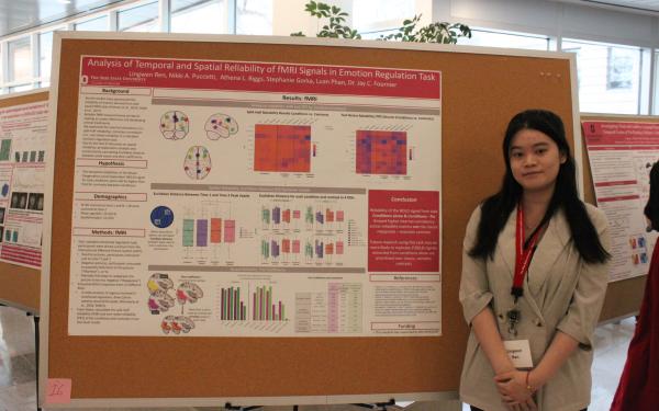
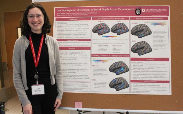
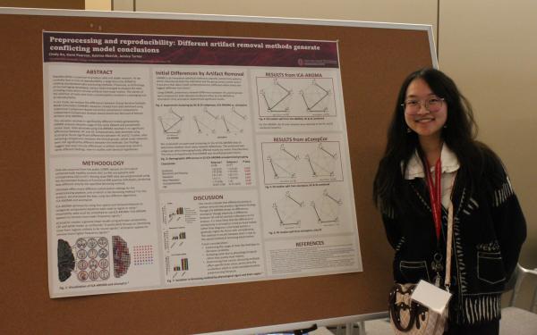
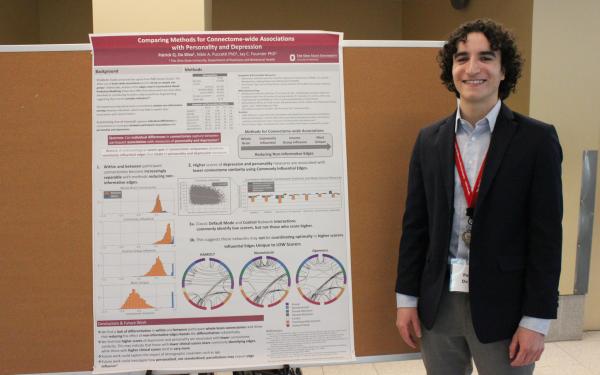
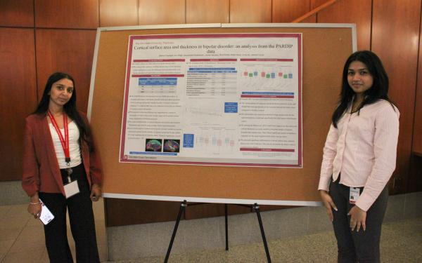
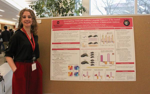

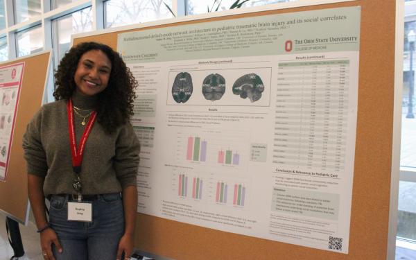
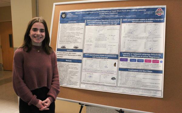
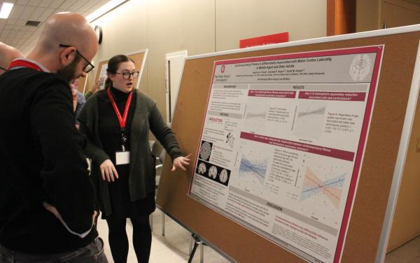
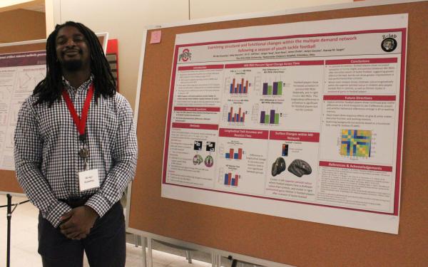
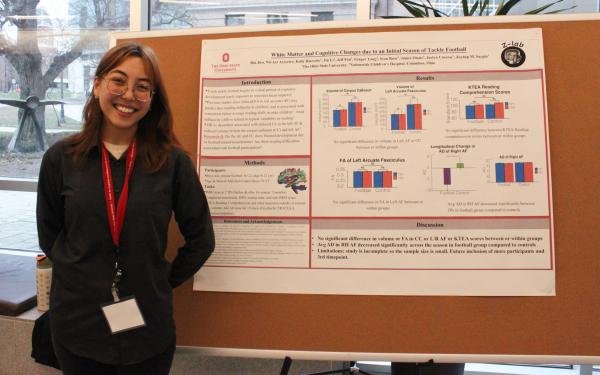


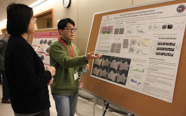
Research Day Committees
Uyen Tram, CCBBI Manager
Adam Gorka, Research Assistant Professor
James Teng, Graduate Research Assistant
Xiangrui Li, CCBBI Assistant Director
Elizabeth Sponseller, MRI Technologist
Ruchika Prakash, CCBBI Director
Kelly Hiersche
Eunjee Ko
Jenny Lin
Dan Zhu
Jay Fournier, Psychiatry and Behavioral Health
Jasmeet Hayes, Psychology
Ruchika Prakash, Psychology
Zeynep Saygin, Psychology
Adam Gorka, Psychology
Julie Golomb, Psychology
Hsin-Hung Li, Psychology
Andrew Leber, Psychology
Scott Hayes, Psychology
David Osher, Psychology
Nicole Puccetti, Psychiatry
Jessica Turner, Psychiatry
Dylan Wagner, Psychology
Baldwin Way, Psychology
