The Imager Spring 2017
The Center for Cognitive and Behavioral Brain Imaging (CCBBI) is a preeminent brain imaging research facility, uniquely designed as an active learning environment. The center provides an innovative, hands-on learning opportunity for Ohio State undergraduate and graduate students. As laboratory research assistants, students are able to help conduct actual research studies alongside world renowned faculty. Undergraduate and graduate students are able to take university credit courses in fMRI taught by brain imaging faculty experts.
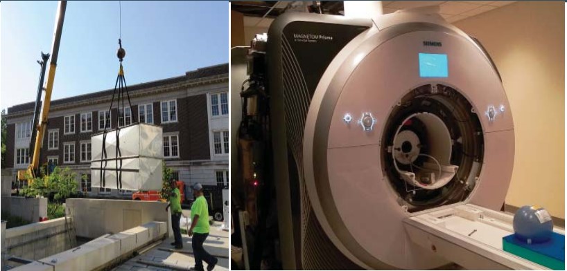
Zack Dunnells, a neuroscience and psychology undergraduate student in Professor Zhong-Lin Lu’s Intro to Functional Magnetic Resonance Imaging (fMRI) class shares his thoughts: “I am doing what I am going to do in grad school already as an undergraduate, so this is giving me preparation and insight to see if this is actually what I want to do.” The course provides a general introduction to MRI, including the physiological bases and principals of functional MRI, design and analysis of fMRI experiments, and operation of the Siemens PrismaFit system. Undergraduate students benefi t directly from this fi rsthand experience, as Zack explains, “This is a unique opportunity for me because I get to experience it (brain imaging techniques) and the training well before I am accepted or apply to graduate programs. It gives me the chance to stand out from another applicant.”
Graduate students and staff employees also benefi t by taking fMRI courses. William “Jason” Riggs, an audiologist at Ohio State’s Eye and Ear Institute, took the class to receive specialized training in neuroimaging design, development and brain imaging techniques. In his work, Jason and colleagues study changes in cognition of patients who have hearing loss and how that aff ects their executive functioning including working memory and other types of neural processes.
By integrating functional MRI and neuroimaging into their already established electrophysiology and cognitive research techniques, Jason explains that researchers can “better understand how changes in the auditory system aff ect a patient and how they respond to treatment such as hearing aids and other types of auditory prostheses.” By comparing a patient’s brain activity pre and post treatment, medical professionals are able to “validate that their intervention and subsequent treatment/rehabilitation plan is appropriate and most eff ective for the patient.” The CCBBI is excited to support the pioneering research being done in the Center, and will continue to contribute to training the country’s brightest students and rising stars in the fi eld of functional resonance imaging.
What kinds of events and activities does the group plan and participate in?
There are several imaging techniques used across different departments at Ohio State. Our meetings aim to tap into this existing knowledge, and share it.
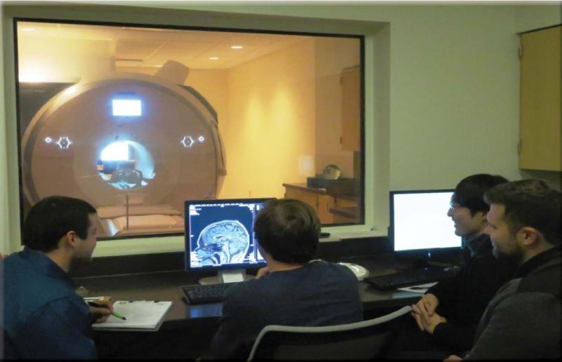
Our student-led meetings provide a forum for students to present on topics such as physiological data, machine learning and new brain imaging methods. Our members also lead hands-on workshops during which students can learn how to work with software packages. This safe environment creates a culture that supports open discussion and methodological critiques at a student level.
Our group organizes and participates in seminars by experts in the fi eld. Last semester, Professor Gary Berntson gave a talk about the logical challenges beginning researchers could face. Most recently, we hosted Professor Rafael Polania from the University of Zurich, who provided a workshop on Transcranial Alternating Current Stimulation. These meetings serve as a great place to learn theories and cutting-edge methods from professionals.
Additionally, we hold group social activities to foster open partnerships between labs and research disciplines, which strengthens interdisciplinary collaborations within the brain imaging community.
How does involvement in the group enrich the academic experience of the members?
From an academic standpoint, the majority of MRI techniques are not learned in the classroom. MRI techniques are largely unique to the specialized, rigorous and rapidly changing methods. In-lab training is our primary source of knowledge, passed down from both advisors and graduate students. Our group provides a common space where developing researchers can explore new methods in a supportive, out-of-lab environment. Our group is open to all levels of expertise, from post-doctoral scientists to first-year grad students — even a few daring research assistants!
lf you would like more information about the CCBBI Student Group and how to join, please visit ccbbistudentgroup.wordpress.com/about.

Orofacial clefts are one of the most common birth defects, affecting approximately 7,000 babies born in the U.S. annually.
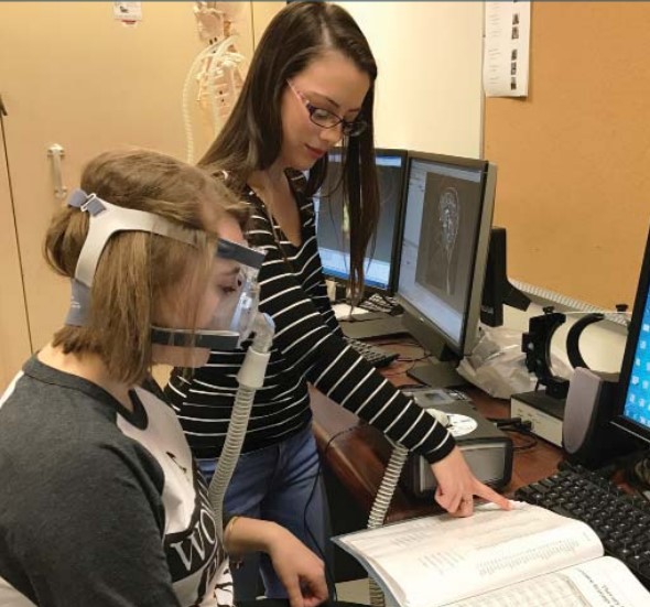
Cleft palate refers to an opening along the roof of the mouth, which can potentially cause many issues related to feeding, hearing, dentition, and speech/resonance in affected individuals. It is estimated that over 20 percent of individuals born with cleft palate continue to experience persisting hypernasality after palatal repair surgery. Hypernasality is a speech resonance disorder in which too much sound and acoustic energy resonate through the nasal cavities, resulting in the deterioration of speech intelligibility. This persisting speech resonance problem may have negative impact on the affected individual’s communication capacity as well as psychosocial well-being.
Please tell us about the research you are conducting at CCBBI.
My research primarily focuses on this subsystem of the speech production mechanism, including the soft palate and surrounding pharyngeal walls (i.e., velopharyngeal mechanism) in the throat region, and aims to identify vital anatomic features of the velopharynx that can serve as reliable prognostic indicators of the speech outcome in individuals with a history of cleft palate. Individuals with a history of cleft palate commonly exhibit structural and functional deviations of the underlying velopharyngeal muscles; nonetheless, the full spectrum of clinical manifestations is not well understood. High-resolution MRI data allow us to quantify various structural parameters, which has potential diagnostic value in patients presenting persisting resonance problems. Our team at the Ohio State Voice/Resonance Lab works toward the goal of identifying key structural parameters that need to be preserved or reconstructed during surgery for better speech outcome. We also use MRI data to gauge morphologic changes induced by an 8-week intensive velopharyngeal closure muscle strengthening program as an alternative behavioral intervention option for treating hypernasality.
What is the impact of your research for the future?
While physical management (i.e., additional surgery) is considered the first-line treatment option for patients with persistent hypernasality, non-surgical behavioral intervention has also been demonstrated to be effective in reducing hypernasality. This will particularly benefit patients who wish to forgo surgery, or those for whom surgery is not a viable option. Our work will lead us to a better understanding of the velopharyngeal structural and functional capacity to be altered through a resistance exercise/muscle strengthening program, which will make an important contribution to quality management of individuals born with cleft palate.
Arkady Konovalov, a graduate student in economics, received a 2015 CCBBI Neuroimaging Award. This scholarship is made possible through the generosity of private donations made to support undergraduate and graduate student researchers in the Center
I work in the fascinating new fi eld of neuroeconomics that combines research questions and methods of economics, cognitive psychology, and neuroscience. Neuroimaging techniques allow us to provide new, important insights into human behavior in general, and reasoning processes, in particular. In economics, there is an increasing interest in studying the intricacies of decision making using non-choice data such as fMRI, and I am very excited to be a part of these new developments.
My project studies how the brain identifies patterns in sequences of events. For scientists, discovering regularities in sometimes seemingly random data is a big part of the job. However, every person learns some types of patterns in their daily lives: it can be traffic, their friends’ habits, or local weather. We already know that the brain can identify patterns even when the person does not realize that a pattern is present in a sequence of events they observe (for example, strings of letters). This unconscious decoding of signals is called “implicit learning.”
The research we conduct in Professor Ian Krajbich’s Neuroeconomics Lab, in the Department of Psychology, focuses on explicit learning. In explicit learning, the individual is purposely learning and is aware of what they are learning. One of the concerns in studying patterns in data is biases: sometimes we see patterns even when there is no real relationship and the outcome was a result of pure chance. This has been a huge problem for data-driven scientific methods for decades. One of the interesting goals of our project is to identify the brain activity associated with these false insights.
In our studies in the CCBBI, participants see a sequence of images and have to respond with a specific button press for each image they observe. If there is a pattern in the sequence, they can respond faster, without visually processing the image, and receive a larger reward. By analyzing their response times and functional imaging data acquired during the scan, we can apply a mathematical model to identify that the brain already has some knowledge about the type of patterns it can encounter, and how it uses an efficient reasoning process to figure out what is going on in the sequence.
While prior studies have identified the main regions in the brain that are generally involved in sequence learning, our research enables us to further distinguish regions that track knowledge about the events in the sequence and knowledge about the pattern itself. We hope that this knowledge will provide a better understanding of how the reasoning process works within specific brain networks: for instance, regions in which activity increases or decreases during pattern learning, and how connectivity between these regions mediates the process.
As the demand for interdisciplinary scholars that can combine methods of neuroscience and economics steadily rises, I am able to greatly further my professional development by being able to practice this research in the CCBBI.
If you would like to learn more about how you can support student research at the CCBBI, please contact: Tammy Parker, Director of Development | parker.465@osu.edu | 614-688-5660
The Siemens Magnetom 3T PrismaFit system upgrade is vital, ensuring that the Center continues to offer the most powerful and state-of-the-art imaging technology to researchers.